 Movie
Movie Controller

[English] 日本語
 Yorodumi
- PDB-6zip: Crystal Structure of Two-Domain Laccase mutant R240A from Strepto... -
+ Open data

- Basic information
| Entry | Database: PDB / ID: 6zip | |||||||||
|---|---|---|---|---|---|---|---|---|---|---|
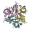
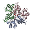
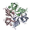
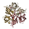
| Title | Crystal Structure of Two-Domain Laccase mutant R240A from Streptomyces griseoflavus | |||||||||
 Components Components | Two-domain laccase | |||||||||
 Keywords Keywords | OXIDOREDUCTASE / Two-Domain Laccase / laccase / Streptomyces griseoflavus | |||||||||
| Function / homology |  Function and homology information Function and homology informationhydroquinone:oxygen oxidoreductase activity / laccase / iron ion transport / copper ion binding / plasma membrane Similarity search - Function | |||||||||
| Biological species |  Streptomyces griseoflavus (bacteria) Streptomyces griseoflavus (bacteria) | |||||||||
| Method |  X-RAY DIFFRACTION / SYNCHROTRON / MOLECULAR REPLACEMENT / Resolution: 2.05 Å X-RAY DIFFRACTION / SYNCHROTRON / MOLECULAR REPLACEMENT / Resolution: 2.05 Å | |||||||||
 Authors Authors | Gabdulkhakov, A.G. / Tishchenko, T.V. / Kolyadenko, I.A. | |||||||||
| Funding support |  Russian Federation, 2items Russian Federation, 2items
| |||||||||
 Citation Citation | Journal: J.Biomol.Struct.Dyn. / Year: 2022 Title: The role of positive charged residue in the proton-transfer mechanism of two-domain laccase from Streptomyces griseoflavus Ac-993. Authors: Gabdulkhakov, A. / Kolyadenko, I. / Oliveira, P. / Tamagnini, P. / Mikhaylina, A. / Tishchenko, S. | |||||||||
| History |
|
- Structure visualization
| Structure viewer | Molecule: MolmilJmol/JSmol |
|---|
- Downloads & links
-Download
| PDBx/mmCIF format | 6zip.cif.gz | 661.7 KB | Display | PDBx/mmCIF format |
|---|---|---|---|---|
| PDB format | pdb6zip.ent.gz | 533.4 KB | Display | PDB format |
| PDBx/mmJSON format | 6zip.json.gz | Tree view | PDBx/mmJSON format | |
| Others |  Other downloads Other downloads |
-Validation report
| Arichive directory | https://data.pdbj.org/pub/pdb/validation_reports/zi/6zipftp://data.pdbj.org/pub/pdb/validation_reports/zi/6zip | HTTPS FTP |
|---|
-Related structure data
| Related structure data |  6zijC  5lhlS C: citing same article ( S: Starting model for refinement |
|---|---|
| Similar structure data |


-Links
 PDBj
PDBj


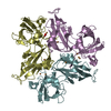
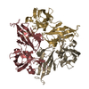
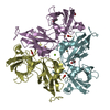
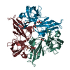
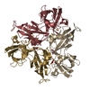
- Assembly
| Deposited unit | 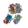
| ||||||||||||
|---|---|---|---|---|---|---|---|---|---|---|---|---|---|
| 1 | 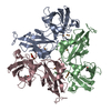
| ||||||||||||
| 2 | 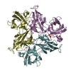
| ||||||||||||
| 3 | 
| ||||||||||||
| 4 | 
| ||||||||||||
| Unit cell |
|
-Components
-Protein , 1 types, 12 molecules ABCDEFGHIJKL
| #1: Protein | Mass: 34665.523 Da / Num. of mol.: 12 / Mutation: R240A Source method: isolated from a genetically manipulated source Source: (gene. exp.) Streptomyces griseoflavus (bacteria) / Production host: |
|---|
-Non-polymers , 5 types, 847 molecules 


| #2: Chemical | ChemComp-CU /  Mass: 63.546 Da / Num. of mol.: 48 / Source method: obtained synthetically / Formula: Cu / Feature type: SUBJECT OF INVESTIGATION Mass: 63.546 Da / Num. of mol.: 48 / Source method: obtained synthetically / Formula: Cu / Feature type: SUBJECT OF INVESTIGATION#3: Chemical |  Mass: 92.094 Da / Num. of mol.: 3 / Source method: obtained synthetically / Formula: C3H8O3 Mass: 92.094 Da / Num. of mol.: 3 / Source method: obtained synthetically / Formula: C3H8O3#4: Chemical |  Mass: 31.999 Da / Num. of mol.: 3 / Source method: obtained synthetically / Formula: O2 / Feature type: SUBJECT OF INVESTIGATION Mass: 31.999 Da / Num. of mol.: 3 / Source method: obtained synthetically / Formula: O2 / Feature type: SUBJECT OF INVESTIGATION#5: Chemical | ChemComp-PER / |  Mass: 31.999 Da / Num. of mol.: 1 / Source method: obtained synthetically / Formula: O2 / Feature type: SUBJECT OF INVESTIGATION Mass: 31.999 Da / Num. of mol.: 1 / Source method: obtained synthetically / Formula: O2 / Feature type: SUBJECT OF INVESTIGATION#6: Water | ChemComp-HOH / | Mass: 18.015 Da / Num. of mol.: 792 / Source method: isolated from a natural source / Formula: H2O |
|---|
-Details
| Has ligand of interest | Y |
|---|
-Experimental details
-Experiment
| Experiment | Method: X-RAY DIFFRACTION / Number of used crystals: 1 |
|---|
- Sample preparation
| Crystal | Density Matthews: 2.03 Å3/Da / Density % sol: 39.53 % |
|---|---|
| Crystal grow | Temperature: 285 K / Method: vapor diffusion, hanging drop / pH: 9.3 / Details: 20 % v/v PEG Smear High 0.1 M Bicine, pH 9.3 |
-Data collection
| Diffraction | Mean temperature: 100 K / Serial crystal experiment: N |
|---|---|
| Diffraction source | Source: SYNCHROTRON / Site: BESSY  / Beamline: 14.1 / Wavelength: 0.861 Å / Beamline: 14.1 / Wavelength: 0.861 Å |
| Detector | Type: DECTRIS PILATUS3 S 6M / Detector: PIXEL / Date: Mar 23, 2019 |
| Radiation | Protocol: SINGLE WAVELENGTH / Monochromatic (M) / Laue (L): M / Scattering type: x-ray |
| Radiation wavelength | Wavelength: 0.861 Å / Relative weight: 1 |
| Reflection | Resolution: 2.05→50 Å / Num. obs: 200840 / % possible obs: 97.6 % / Redundancy: 2.67 % / Biso Wilson estimate: 35.33 Å2 / CC1/2: 0.99 / Rmerge(I) obs: 0.085 / Net I/σ(I): 7.09 |
| Reflection shell | Resolution: 2.05→2.1 Å / Rmerge(I) obs: 0.72 / Mean I/σ(I) obs: 1.28 / Num. unique obs: 14801 / CC1/2: 0.69 / % possible all: 97.2 |
- Processing
| Software |
| |||||||||||||||||||||||||||||||||||||||||||||||||||||||||||||||||||||||||||||||||||||||||||||||||||||||||||||||||||||||||||||||||||||||||||||||||||||||||||||||||||||||||||||||||||||||||||||||||||||||||||||||||||||||||
|---|---|---|---|---|---|---|---|---|---|---|---|---|---|---|---|---|---|---|---|---|---|---|---|---|---|---|---|---|---|---|---|---|---|---|---|---|---|---|---|---|---|---|---|---|---|---|---|---|---|---|---|---|---|---|---|---|---|---|---|---|---|---|---|---|---|---|---|---|---|---|---|---|---|---|---|---|---|---|---|---|---|---|---|---|---|---|---|---|---|---|---|---|---|---|---|---|---|---|---|---|---|---|---|---|---|---|---|---|---|---|---|---|---|---|---|---|---|---|---|---|---|---|---|---|---|---|---|---|---|---|---|---|---|---|---|---|---|---|---|---|---|---|---|---|---|---|---|---|---|---|---|---|---|---|---|---|---|---|---|---|---|---|---|---|---|---|---|---|---|---|---|---|---|---|---|---|---|---|---|---|---|---|---|---|---|---|---|---|---|---|---|---|---|---|---|---|---|---|---|---|---|---|---|---|---|---|---|---|---|---|---|---|---|---|---|---|---|---|
| Refinement | Method to determine structure: MOLECULAR REPLACEMENT Starting model: 5LHL Resolution: 2.05→47.4 Å / SU ML: 0.2382 / Cross valid method: FREE R-VALUE / σ(F): 1.97 / Phase error: 23.6624 Stereochemistry target values: GeoStd + Monomer Library + CDL v1.2
| |||||||||||||||||||||||||||||||||||||||||||||||||||||||||||||||||||||||||||||||||||||||||||||||||||||||||||||||||||||||||||||||||||||||||||||||||||||||||||||||||||||||||||||||||||||||||||||||||||||||||||||||||||||||||
| Solvent computation | Shrinkage radii: 0.9 Å / VDW probe radii: 1.11 Å / Solvent model: FLAT BULK SOLVENT MODEL | |||||||||||||||||||||||||||||||||||||||||||||||||||||||||||||||||||||||||||||||||||||||||||||||||||||||||||||||||||||||||||||||||||||||||||||||||||||||||||||||||||||||||||||||||||||||||||||||||||||||||||||||||||||||||
| Displacement parameters | Biso mean: 40.55 Å2 | |||||||||||||||||||||||||||||||||||||||||||||||||||||||||||||||||||||||||||||||||||||||||||||||||||||||||||||||||||||||||||||||||||||||||||||||||||||||||||||||||||||||||||||||||||||||||||||||||||||||||||||||||||||||||
| Refinement step | Cycle: LAST / Resolution: 2.05→47.4 Å
| |||||||||||||||||||||||||||||||||||||||||||||||||||||||||||||||||||||||||||||||||||||||||||||||||||||||||||||||||||||||||||||||||||||||||||||||||||||||||||||||||||||||||||||||||||||||||||||||||||||||||||||||||||||||||
| Refine LS restraints |
| |||||||||||||||||||||||||||||||||||||||||||||||||||||||||||||||||||||||||||||||||||||||||||||||||||||||||||||||||||||||||||||||||||||||||||||||||||||||||||||||||||||||||||||||||||||||||||||||||||||||||||||||||||||||||
| LS refinement shell |
|
