 Movie
Movie Controller


+ Open data

- Basic information
| Entry | Database: PDB / ID: 7bk7 | ||||||
|---|---|---|---|---|---|---|---|
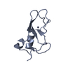
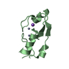
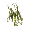
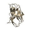
| Title | PfCopC mutant - D83N | ||||||
 Components Components | Putative copper resistance protein | ||||||
 Keywords Keywords | METAL BINDING PROTEIN / Copper binding / copper transport | ||||||
| Function / homology |  Function and homology information Function and homology informationcopper ion transport / response to copper ion / periplasmic space / copper ion binding / plasma membrane Similarity search - Function | ||||||
| Biological species |  Pseudomonas fluorescens (bacteria) Pseudomonas fluorescens (bacteria) | ||||||
| Method |  X-RAY DIFFRACTION / SYNCHROTRON / MOLECULAR REPLACEMENT / Resolution: 2.3 Å X-RAY DIFFRACTION / SYNCHROTRON / MOLECULAR REPLACEMENT / Resolution: 2.3 Å | ||||||
 Authors Authors | Muderspach, S.J. / Ipsen, J. / Rollan, C.H. / Bertelsen, A.B. / Norholm, M.H.H. / Johansen, K.S. / Lo Leggio, L. | ||||||
| Funding support |  Denmark, 1items Denmark, 1items
| ||||||
 Citation Citation | Journal: Febs Lett. / Year: 2021 Title: Copper binding and reactivity at the histidine brace motif: insights from mutational analysis of the Pseudomonas fluorescens copper chaperone CopC. Authors: Ipsen, J.O. / Hernandez-Rollan, C. / Muderspach, S.J. / Brander, S. / Bertelsen, A.B. / Jensen, P.E. / Norholm, M.H.H. / Lo Leggio, L. / Johansen, K.S. | ||||||
| History |
|
- Structure visualization
| Structure viewer | Molecule: MolmilJmol/JSmol |
|---|
- Downloads & links
-Download
| PDBx/mmCIF format | 7bk7.cif.gz | 55.2 KB | Display | PDBx/mmCIF format |
|---|---|---|---|---|
| PDB format | pdb7bk7.ent.gz | Display | PDB format | |
| PDBx/mmJSON format | 7bk7.json.gz | Tree view | PDBx/mmJSON format | |
| Others |  Other downloads Other downloads |
-Validation report
| Summary document | 7bk7_validation.pdf.gz | 451.4 KB | Display | wwPDB validaton report |
|---|---|---|---|---|
| Full document | 7bk7_full_validation.pdf.gz | 455 KB | Display | |
| Data in XML | 7bk7_validation.xml.gz | 10.7 KB | Display | |
| Data in CIF | 7bk7_validation.cif.gz | 13.8 KB | Display | |
| Arichive directory | https://data.pdbj.org/pub/pdb/validation_reports/bk/7bk7ftp://data.pdbj.org/pub/pdb/validation_reports/bk/7bk7 | HTTPS FTP |
-Related structure data
| Related structure data |  7bk5C  7bk6C  6nfqS C: citing same article ( S: Starting model for refinement |
|---|---|
| Similar structure data |


-Links
 PDBj
PDBj- Assembly
| Deposited unit | 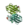
| ||||||||
|---|---|---|---|---|---|---|---|---|---|
| 1 | 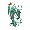
| ||||||||
| 2 | 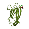
| ||||||||
| Unit cell |
|
-Components
-Protein , 1 types, 2 molecules AAABBB
| #1: Protein | Mass: 10109.310 Da / Num. of mol.: 2 / Mutation: D83N Source method: isolated from a genetically manipulated source Source: (gene. exp.) Pseudomonas fluorescens (bacteria) / Strain: SBW25 / Gene: PFLU_3946Production host: References: UniProt: C3JYL7 |
|---|
-Non-polymers , 5 types, 81 molecules 


| #2: Chemical |  Mass: 59.044 Da / Num. of mol.: 2 / Source method: obtained synthetically / Formula: C2H3O2 Mass: 59.044 Da / Num. of mol.: 2 / Source method: obtained synthetically / Formula: C2H3O2#3: Chemical | ChemComp-CU / |  Mass: 63.546 Da / Num. of mol.: 1 / Source method: obtained synthetically / Formula: Cu Mass: 63.546 Da / Num. of mol.: 1 / Source method: obtained synthetically / Formula: Cu#4: Chemical |  Mass: 35.453 Da / Num. of mol.: 3 / Source method: obtained synthetically / Formula: Cl Mass: 35.453 Da / Num. of mol.: 3 / Source method: obtained synthetically / Formula: Cl#5: Chemical | ChemComp-SO4 / |  Mass: 96.063 Da / Num. of mol.: 1 / Source method: obtained synthetically / Formula: SO4 Mass: 96.063 Da / Num. of mol.: 1 / Source method: obtained synthetically / Formula: SO4#6: Water | ChemComp-HOH / | Mass: 18.015 Da / Num. of mol.: 74 / Source method: isolated from a natural source / Formula: H2O |
|---|
-Details
| Has ligand of interest | N |
|---|
-Experimental details
-Experiment
| Experiment | Method: X-RAY DIFFRACTION / Number of used crystals: 1 |
|---|
- Sample preparation
| Crystal | Density Matthews: 3.58 Å3/Da / Density % sol: 65.68 % |
|---|---|
| Crystal grow | Temperature: 298 K / Method: vapor diffusion, sitting drop / pH: 4.6 / Details: 2.9 M ammonium sulphate, 0.1 M sodium acetate |
-Data collection
| Diffraction | Mean temperature: 100 K / Serial crystal experiment: N |
|---|---|
| Diffraction source | Source: SYNCHROTRON / Site: ESRF  / Beamline: ID23-2 / Wavelength: 0.87313 Å / Beamline: ID23-2 / Wavelength: 0.87313 Å |
| Detector | Type: DECTRIS PILATUS3 X 2M / Detector: PIXEL / Date: Oct 7, 2020 |
| Radiation | Protocol: SINGLE WAVELENGTH / Monochromatic (M) / Laue (L): M / Scattering type: x-ray |
| Radiation wavelength | Wavelength: 0.87313 Å / Relative weight: 1 |
| Reflection | Resolution: 2.3→50 Å / Num. obs: 12725 / % possible obs: 99.8 % / Redundancy: 5.64 % / CC1/2: 0.998 / Rrim(I) all: 0.102 / Net I/σ(I): 10.52 |
| Reflection shell | Resolution: 2.3→2.36 Å / Num. unique obs: 1884 / CC1/2: 0.457 / Rrim(I) all: 1.962 |
- Processing
| Software |
| |||||||||||||||||||||||||||||||||||||||||||||||||||||||||||||||||||||||||||||||||||||||||||||||||||||||||||||||||||||||||||||||||||||||||||||||||||||||||||
|---|---|---|---|---|---|---|---|---|---|---|---|---|---|---|---|---|---|---|---|---|---|---|---|---|---|---|---|---|---|---|---|---|---|---|---|---|---|---|---|---|---|---|---|---|---|---|---|---|---|---|---|---|---|---|---|---|---|---|---|---|---|---|---|---|---|---|---|---|---|---|---|---|---|---|---|---|---|---|---|---|---|---|---|---|---|---|---|---|---|---|---|---|---|---|---|---|---|---|---|---|---|---|---|---|---|---|---|---|---|---|---|---|---|---|---|---|---|---|---|---|---|---|---|---|---|---|---|---|---|---|---|---|---|---|---|---|---|---|---|---|---|---|---|---|---|---|---|---|---|---|---|---|---|---|---|---|
| Refinement | Method to determine structure: MOLECULAR REPLACEMENT Starting model: 6NFQ Resolution: 2.3→35.266 Å / Cor.coef. Fo:Fc: 0.962 / Cor.coef. Fo:Fc free: 0.938 / WRfactor Rfree: 0.289 / WRfactor Rwork: 0.22 / SU B: 8.819 / SU ML: 0.211 / Average fsc free: 0.8515 / Average fsc work: 0.8713 / Cross valid method: FREE R-VALUE / ESU R: 0.255 / ESU R Free: 0.226 Details: Hydrogens have been added in their riding positions
| |||||||||||||||||||||||||||||||||||||||||||||||||||||||||||||||||||||||||||||||||||||||||||||||||||||||||||||||||||||||||||||||||||||||||||||||||||||||||||
| Solvent computation | Ion probe radii: 0.8 Å / Shrinkage radii: 0.8 Å / VDW probe radii: 1.2 Å / Solvent model: MASK BULK SOLVENT | |||||||||||||||||||||||||||||||||||||||||||||||||||||||||||||||||||||||||||||||||||||||||||||||||||||||||||||||||||||||||||||||||||||||||||||||||||||||||||
| Displacement parameters | Biso mean: 70.442 Å2
| |||||||||||||||||||||||||||||||||||||||||||||||||||||||||||||||||||||||||||||||||||||||||||||||||||||||||||||||||||||||||||||||||||||||||||||||||||||||||||
| Refinement step | Cycle: LAST / Resolution: 2.3→35.266 Å
| |||||||||||||||||||||||||||||||||||||||||||||||||||||||||||||||||||||||||||||||||||||||||||||||||||||||||||||||||||||||||||||||||||||||||||||||||||||||||||
| Refine LS restraints |
| |||||||||||||||||||||||||||||||||||||||||||||||||||||||||||||||||||||||||||||||||||||||||||||||||||||||||||||||||||||||||||||||||||||||||||||||||||||||||||
| LS refinement shell |
|
