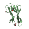
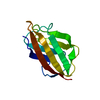
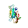
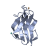
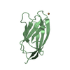
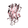
[English] 日本語
Yorodumi
- PDB-6nfq: CopC from Pseudomonas fluorescens -

+
Open data


ID or keywords:

Loading...

-
Basic information

Entry
Database: PDB / ID: 6nfq
TitleCopC from Pseudomonas fluorescens
ComponentsCopC
KeywordsMETAL BINDING PROTEIN / CopC / metallochaperone / copper binding
Function / homology
Function and homology information


copper ion transport / response to copper ion / periplasmic space / copper ion binding / plasma membrane
Similarity search - Function
Copper transport protein C/D / : / CopC domain / CopC domain / Copper resistance protein CopC/internalin, immunoglobulin-like / Immunoglobulin E-set
Similarity search - Domain/homology
COPPER (II) ION / YTTRIUM (III) ION / Copper resistance protein C
Similarity search - Component
Biological speciesPseudomonas fluorescens (bacteria)
MethodX-RAY DIFFRACTION / SYNCHROTRON / MOLECULAR REPLACEMENT / Resolution: 2 Å
AuthorsMaher, M.J.
Funding support Australia, 2items
OrganizationGrant numberCountry
Australian Research Council (ARC)DP140102746 Australia
Australian Research Council (ARC)DP130100728 Australia
CitationJournal: J. Inorg. Biochem. / Year: 2019
Title: The crystal structure of the CopC protein from Pseudomonas fluorescens reveals amended classifications for the CopC protein family.
Authors: Udagedara, S.R. / Wijekoon, C.J.K. / Xiao, Z. / Wedd, A.G. / Maher, M.J.
History
DepositionDec 20, 2018Deposition site: RCSB / Processing site: RCSB
Revision 1.0Apr 24, 2019Provider: repository / Type: Initial release
Revision 1.1Jan 1, 2020Group: Author supporting evidence / Category: pdbx_audit_support / Item: _pdbx_audit_support.funding_organization
Revision 1.2Oct 11, 2023Group: Data collection / Database references ...Data collection / Database references / Derived calculations / Refinement description
Category: chem_comp_atom / chem_comp_bond ...chem_comp_atom / chem_comp_bond / database_2 / pdbx_initial_refinement_model / pdbx_struct_conn_angle / struct_conn
Item: _database_2.pdbx_DOI / _database_2.pdbx_database_accession ..._database_2.pdbx_DOI / _database_2.pdbx_database_accession / _pdbx_struct_conn_angle.ptnr1_auth_comp_id / _pdbx_struct_conn_angle.ptnr1_auth_seq_id / _pdbx_struct_conn_angle.ptnr1_label_asym_id / _pdbx_struct_conn_angle.ptnr1_label_atom_id / _pdbx_struct_conn_angle.ptnr1_label_comp_id / _pdbx_struct_conn_angle.ptnr1_label_seq_id / _pdbx_struct_conn_angle.ptnr3_auth_comp_id / _pdbx_struct_conn_angle.ptnr3_auth_seq_id / _pdbx_struct_conn_angle.ptnr3_label_asym_id / _pdbx_struct_conn_angle.ptnr3_label_atom_id / _pdbx_struct_conn_angle.ptnr3_label_comp_id / _pdbx_struct_conn_angle.ptnr3_label_seq_id / _pdbx_struct_conn_angle.value / _struct_conn.pdbx_dist_value / _struct_conn.ptnr1_auth_asym_id / _struct_conn.ptnr1_auth_comp_id / _struct_conn.ptnr1_auth_seq_id / _struct_conn.ptnr1_label_asym_id / _struct_conn.ptnr1_label_atom_id / _struct_conn.ptnr1_label_comp_id / _struct_conn.ptnr1_label_seq_id / _struct_conn.ptnr2_auth_asym_id / _struct_conn.ptnr2_auth_comp_id / _struct_conn.ptnr2_auth_seq_id / _struct_conn.ptnr2_label_asym_id / _struct_conn.ptnr2_label_atom_id / _struct_conn.ptnr2_label_comp_id / _struct_conn.ptnr2_symmetry

-
Structure visualization

Structure viewerMolecule:
MolmilJmol/JSmol

Downloads & links

-
Assembly

Deposited unit
A: CopC
B: CopC
C: CopC
hetero molecules


Theoretical massNumber of molelcules
Total (without water)38,2047
Polymers37,9253
Non-polymers2804
Water1,72996
1
A: CopC
hetero molecules


Theoretical massNumber of molelcules
Total (without water)12,7943
Polymers12,6421
Non-polymers1522
Water181
TypeNameSymmetry operationNumber
identity operation1_555x,y,z1
2
B: CopC
hetero molecules


Theoretical massNumber of molelcules
Total (without water)12,7052
Polymers12,6421
Non-polymers641
Water181
TypeNameSymmetry operationNumber
identity operation1_555x,y,z1
3
C: CopC
hetero molecules


Theoretical massNumber of molelcules
Total (without water)12,7052
Polymers12,6421
Non-polymers641
Water181
TypeNameSymmetry operationNumber
identity operation1_555x,y,z1
Unit cell
Length a, b, c (Å)77.730, 111.319, 82.222
Angle α, β, γ (deg.)90.00, 90.00, 90.00
Int Tables number20
Space group name H-MC2221
Components on special symmetry positions
IDModelComponents
11A-202-

YT3

-
Components

#1: Protein CopC


Mass: 12641.553 Da / Num. of mol.: 3
Source method: isolated from a genetically manipulated source
Source: (gene. exp.) Pseudomonas fluorescens (bacteria) / Production host: Escherichia coli (E. coli) / References: UniProt: C3JYL7*PLUS
#2: Chemical ChemComp-CU / COPPER (II) ION


Mass: 63.546 Da / Num. of mol.: 3 / Source method: obtained synthetically / Formula: Cu
#3: Chemical ChemComp-YT3 / YTTRIUM (III) ION


Mass: 88.906 Da / Num. of mol.: 1 / Source method: obtained synthetically / Formula: Y
#4: Water ChemComp-HOH / water


Mass: 18.015 Da / Num. of mol.: 96 / Source method: isolated from a natural source / Formula: H2O

-
Experimental details

-
Experiment

ExperimentMethod: X-RAY DIFFRACTION / Number of used crystals: 1

-
Sample preparation

CrystalDensity Matthews: 2.34 Å3/Da / Density % sol: 47.55 %
Crystal growTemperature: 293 K / Method: vapor diffusion, hanging drop / pH: 6.7
Details: 2.5 M ammonium sulfate, 0.1 M Bis-Tris propane, pH 6.7, 2 mM yttrium chloride hexahydrate

-
Data collection

DiffractionMean temperature: 100 K / Serial crystal experiment: N
Diffraction sourceSource: SYNCHROTRON / Site: Australian Synchrotron / Beamline: MX2 / Wavelength: 0.9537 Å
DetectorType: ADSC QUANTUM 315r / Detector: CCD / Date: Apr 1, 2015
RadiationProtocol: SINGLE WAVELENGTH / Monochromatic (M) / Laue (L): M / Scattering type: x-ray
Radiation wavelengthWavelength: 0.9537 Å / Relative weight: 1
ReflectionResolution: 2→46 Å / Num. obs: 23030 / % possible obs: 99.5 % / Redundancy: 6 % / Net I/σ(I): 9.7
Reflection shellResolution: 2→2.05 Å

-
Processing

Software
NameVersionClassification
HKL-20005.8.0230data reduction
SCALEPACKdata scaling
PHASERphasing
REFMACrefinement
RefinementMethod to determine structure: MOLECULAR REPLACEMENT
Starting model: PDB entry 2C9Q
Resolution: 2→46 Å / Cor.coef. Fo:Fc: 0.95 / Cor.coef. Fo:Fc free: 0.935 / SU B: 3.725 / SU ML: 0.104 / Cross valid method: THROUGHOUT / ESU R: 0.039 / ESU R Free: 0.035 / Details: HYDROGENS HAVE BEEN ADDED IN THE RIDING POSITIONS
RfactorNum. reflection% reflectionSelection details
Rfree0.25988 1238 5.1 %RANDOM
Rwork0.22791 ---
obs0.2296 23030 99.5 %-
Solvent computationIon probe radii: 0.8 Å / Shrinkage radii: 0.8 Å / VDW probe radii: 1.2 Å
Displacement parametersBiso mean: 39.18 Å2
Baniso -1Baniso -2Baniso -3
1-31.11 Å20 Å20 Å2
2---11.18 Å20 Å2
3----19.93 Å2
Refinement stepCycle: 1 / Resolution: 2→46 Å
ProteinNucleic acidLigandSolventTotal
Num. atoms2122 0 4 96 2222
Refine LS restraints
Refine-IDTypeDev idealDev ideal targetNumber
X-RAY DIFFRACTIONr_bond_refined_d0.010.0142164
X-RAY DIFFRACTIONr_bond_other_d0.0010.0172029
X-RAY DIFFRACTIONr_angle_refined_deg1.3671.6592950
X-RAY DIFFRACTIONr_angle_other_deg0.8691.6544729
X-RAY DIFFRACTIONr_dihedral_angle_1_deg7.1675286
X-RAY DIFFRACTIONr_dihedral_angle_2_deg39.52624.875
X-RAY DIFFRACTIONr_dihedral_angle_3_deg16.64515340
X-RAY DIFFRACTIONr_dihedral_angle_4_deg20.394153
X-RAY DIFFRACTIONr_chiral_restr0.0630.2307
X-RAY DIFFRACTIONr_gen_planes_refined0.0070.022393
X-RAY DIFFRACTIONr_gen_planes_other0.0010.02379
X-RAY DIFFRACTIONr_nbd_refined
X-RAY DIFFRACTIONr_nbd_other
X-RAY DIFFRACTIONr_nbtor_refined
X-RAY DIFFRACTIONr_nbtor_other
X-RAY DIFFRACTIONr_xyhbond_nbd_refined
X-RAY DIFFRACTIONr_xyhbond_nbd_other
X-RAY DIFFRACTIONr_metal_ion_refined
X-RAY DIFFRACTIONr_metal_ion_other
X-RAY DIFFRACTIONr_symmetry_vdw_refined
X-RAY DIFFRACTIONr_symmetry_vdw_other
X-RAY DIFFRACTIONr_symmetry_hbond_refined
X-RAY DIFFRACTIONr_symmetry_hbond_other
X-RAY DIFFRACTIONr_symmetry_metal_ion_refined
X-RAY DIFFRACTIONr_symmetry_metal_ion_other
X-RAY DIFFRACTIONr_mcbond_it2.9783.7831153
X-RAY DIFFRACTIONr_mcbond_other2.9633.7811152
X-RAY DIFFRACTIONr_mcangle_it3.8545.6621436
X-RAY DIFFRACTIONr_mcangle_other3.8535.6641437
X-RAY DIFFRACTIONr_scbond_it3.6614.161010
X-RAY DIFFRACTIONr_scbond_other3.664.1621011
X-RAY DIFFRACTIONr_scangle_it
X-RAY DIFFRACTIONr_scangle_other5.4026.0671513
X-RAY DIFFRACTIONr_long_range_B_refined6.58745.0472179
X-RAY DIFFRACTIONr_long_range_B_other6.58545.0182172
X-RAY DIFFRACTIONr_rigid_bond_restr
X-RAY DIFFRACTIONr_sphericity_free
X-RAY DIFFRACTIONr_sphericity_bonded
LS refinement shellResolution: 2.002→2.054 Å / Total num. of bins used: 20
RfactorNum. reflection% reflection
Rfree0.415 89 -
Rwork0.323 1659 -
obs--98.2 %

+
About Yorodumi

-
News

-
Feb 9, 2022. New format data for meta-information of EMDB entries

New format data for meta-information of EMDB entries

  • Version 3 of the EMDB header file is now the official format.
  • The previous official version 1.9 will be removed from the archive.

Related info.:EMDB header

External links:wwPDB to switch to version 3 of the EMDB data model

-
Aug 12, 2020. Covid-19 info

Covid-19 info

URL: https://pdbjlvh1.pdbj.org/emnavi/covid19.php

New page: Covid-19 featured information page in EM Navigator.

Related info.:Covid-19 info / Mar 5, 2020. Novel coronavirus structure data

+
Mar 5, 2020. Novel coronavirus structure data

Novel coronavirus structure data

Related info.:Yorodumi Speices / Aug 12, 2020. Covid-19 info

External links:COVID-19 featured content - PDBj / Molecule of the Month (242):Coronavirus Proteases

+
Jan 31, 2019. EMDB accession codes are about to change! (news from PDBe EMDB page)

EMDB accession codes are about to change! (news from PDBe EMDB page)

  • The allocation of 4 digits for EMDB accession codes will soon come to an end. Whilst these codes will remain in use, new EMDB accession codes will include an additional digit and will expand incrementally as the available range of codes is exhausted. The current 4-digit format prefixed with “EMD-” (i.e. EMD-XXXX) will advance to a 5-digit format (i.e. EMD-XXXXX), and so on. It is currently estimated that the 4-digit codes will be depleted around Spring 2019, at which point the 5-digit format will come into force.
  • The EM Navigator/Yorodumi systems omit the EMD- prefix.

Related info.:Q: What is EMD? / ID/Accession-code notation in Yorodumi/EM Navigator

External links:EMDB Accession Codes are Changing Soon! / Contact to PDBj

+
Jul 12, 2017. Major update of PDB

Major update of PDB

  • wwPDB released updated PDB data conforming to the new PDBx/mmCIF dictionary.
  • This is a major update changing the version number from 4 to 5, and with Remediation, in which all the entries are updated.
  • In this update, many items about electron microscopy experimental information are reorganized (e.g. em_software).
  • Now, EM Navigator and Yorodumi are based on the updated data.

External links:wwPDB Remediation / Enriched Model Files Conforming to OneDep Data Standards Now Available in the PDB FTP Archive

-
Yorodumi

Thousand views of thousand structures

  • Yorodumi is a browser for structure data from EMDB, PDB, SASBDB, etc.
  • This page is also the successor to EM Navigator detail page, and also detail information page/front-end page for Omokage search.
  • The word "yorodu" (or yorozu) is an old Japanese word meaning "ten thousand". "mi" (miru) is to see.

Related info.:EMDB / PDB / SASBDB / Comparison of 3 databanks / Yorodumi Search / Aug 31, 2016. New EM Navigator & Yorodumi / Yorodumi Papers / Jmol/JSmol / Function and homology information / Changes in new EM Navigator and Yorodumi

Read more