Mass: 18.015 Da / Num. of mol.: 83 / Source method: isolated from a natural source / Formula: H2O
Compound details
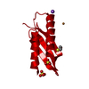
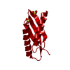
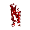
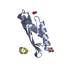
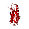
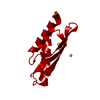
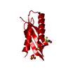
FACILITATES COPPER RESISTANCE BY SEQUESTRATION OF COPPER IN THE PERIPLASM ALONG WITH THE COPPER- ...FACILITATES COPPER RESISTANCE BY SEQUESTRATION OF COPPER IN THE PERIPLASM ALONG WITH THE COPPER-BINDING PROTEIN COPA.
-
Experimental details
-
Experiment
Experiment
Method: X-RAY DIFFRACTION / Number of used crystals: 1
-
Sample preparation
Crystal
Density Matthews: 1.95 Å3/Da / Density % sol: 36.32 %
Crystal grow
pH: 7.5 Details: 2.0 M AMMONIUM SULFATE, 0.1 M SODIUM HEPES, PH 7.5, 2% (W/V) PEG 400
Resolution: 1.6→40 Å / Cor.coef. Fo:Fc: 0.958 / Cor.coef. Fo:Fc free: 0.954 / SU B: 3.297 / SU ML: 0.059 / Cross valid method: THROUGHOUT / ESU R: 0.095 / ESU R Free: 0.097 / Stereochemistry target values: MAXIMUM LIKELIHOOD / Details: HYDROGENS HAVE BEEN ADDED IN THE RIDING POSITIONS.
Rfactor
Num. reflection
% reflection
Selection details
Rfree
0.225
633
4.9 %
RANDOM
Rwork
0.187
-
-
-
obs
0.189
12349
99.4 %
-
Solvent computation
Ion probe radii: 0.8 Å / Shrinkage radii: 0.8 Å / VDW probe radii: 1.2 Å / Solvent model: BABINET MODEL WITH MASK
 Movie
Movie Controller


 Open data
 Basic information
 Components
 Keywords
 Function and homology information
 PSEUDOMONAS SYRINGAE PV. TOMATO (bacteria)
 X-RAY DIFFRACTION /
  Authors
 Citation
 Structure visualization
 Downloads & links
 Other downloads


 PDBj
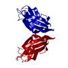
PDBj Assembly
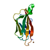

 Mass: 63.546 Da / Num. of mol.: 2 / Source method: obtained synthetically / Formula: Cu
 Mass: 18.015 Da / Num. of mol.: 83 / Source method: isolated from a natural source / Formula: H2O
 Sample preparation
 Processing