 Movie
Movie Controller

[English] 日本語
 Yorodumi
- PDB-7bjp: The cryo-EM structure of vesivirus 2117, an adventitious agent an... -
+ Open data

- Basic information
| Entry | Database: PDB / ID: 7bjp | |||||||||
|---|---|---|---|---|---|---|---|---|---|---|
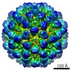
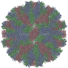
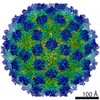
| Title | The cryo-EM structure of vesivirus 2117, an adventitious agent and possible cause of haemorrhagic gastroenteritis in dogs. | |||||||||
 Components Components | Capsid protein | |||||||||
 Keywords Keywords | VIRAL PROTEIN / capsid / calicivirus / vp1 | |||||||||
| Function / homology | T=3 icosahedral viral capsid / Calicivirus coat protein / Calicivirus coat protein / Picornavirus/Calicivirus coat protein / Viral coat protein subunit / host cell cytoplasm / Capsid protein VP1 Function and homology information Function and homology information | |||||||||
| Biological species |  Calicivirus isolate 2117 Calicivirus isolate 2117 | |||||||||
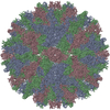
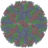
| Method | ELECTRON MICROSCOPY / single particle reconstruction / cryo EM / Resolution: 3.65 Å | |||||||||
 Authors Authors | Sutherland, H. / Conley, M.J. / Emmott, E. / Streetley, J. / Goodfellow, I.G. / Bhella, D. | |||||||||
| Funding support |  United Kingdom, 2items United Kingdom, 2items
| |||||||||
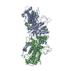
 Citation Citation | Journal: J Virol / Year: 2021 Title: The Cryo-EM Structure of Vesivirus 2117 Highlights Functional Variations in Entry Pathways for Viruses in Different Clades of the Genus. Authors: Hazel Sutherland / Michaela J Conley / Edward Emmott / James Streetley / Ian G Goodfellow / David Bhella / Abstract: Vesivirus 2117 is an adventitious agent that has been responsible for lost productivity in biopharmaceutical production following contamination of Chinese hamster ovary cell cultures in commercial ...Vesivirus 2117 is an adventitious agent that has been responsible for lost productivity in biopharmaceutical production following contamination of Chinese hamster ovary cell cultures in commercial bioreactors. A member of the , 2117 is classified within the Vesivirus genus in a clade that includes canine and mink caliciviruses but is distinct from the vesicular exanthema of swine virus (VESV) clade, which includes the extensively studied feline calicivirus (FCV). We have used cryogenic electron microscopy (cryo-EM) to determine the structure of the capsid of this small, icosahedral, positive-sense-RNA-containing virus. We show that the outer face of the dimeric capsomeres, which contains the receptor binding site and major immunodominant epitopes in all caliciviruses studied thus far, is quite different from that of FCV. This is a consequence of a 22-amino-acid insertion in the sequence of the FCV major capsid protein that forms a "cantilevered arm" that both plays an important role in receptor engagement and undergoes structural rearrangements thought to be important for genome delivery to the cytosol. Our data highlight a potentially important difference in the attachment and entry pathways employed by the different clades of the genus. Vesivirus 2117 has caused significant losses in manufacturing of biopharmaceutical products following contamination of cell cultures used in their production. We report the structure of the vesivirus 2117 capsid, the shell that encloses the virus's genome. Comparison of this structure with that of a related vesivirus, feline calicivirus (FCV), highlighted potentially important differences related to virus attachment and entry. Our findings suggest that these two viruses may bind differently to receptors at the host cell surface. We also show that a region of the capsid protein of FCV that rearranges following receptor engagement is not present in vesivirus 2117. These structural changes in the FCV capsid have been shown to allow the assembly of a portal-like structure that is hypothesized to deliver the viral genome to the cell's interior. Our data suggest that the 2117 portal assembly may employ a different means of anchoring to the outer face of the capsid. #1: Journal: Biorxiv / Year: 2021Title: The cryo-EM structure of vesivirus 2117 highlights functional variations in entry pathways for viruses in different clades of the Vesivirus genus Authors: Sutherland, H. / Conley, M.J. / Emmott, E. / Streetley, J. / Goodfellow, I.G. / Bhella, D. | |||||||||
| History |
|
- Structure visualization
| Movie |
Movie viewer |
|---|---|
| Structure viewer | Molecule: MolmilJmol/JSmol |
- Downloads & links
-Download
| PDBx/mmCIF format | 7bjp.cif.gz | 492.3 KB | Display | PDBx/mmCIF format |
|---|---|---|---|---|
| PDB format | pdb7bjp.ent.gz | 410.2 KB | Display | PDB format |
| PDBx/mmJSON format | 7bjp.json.gz | Tree view | PDBx/mmJSON format | |
| Others |  Other downloads Other downloads |
-Validation report
| Arichive directory | https://data.pdbj.org/pub/pdb/validation_reports/bj/7bjpftp://data.pdbj.org/pub/pdb/validation_reports/bj/7bjp | HTTPS FTP |
|---|
-Related structure data
| Related structure data | 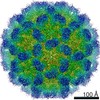 12194MC M: map data used to model this data C: citing same article ( |
|---|---|
| Similar structure data | |
| EM raw data | EMPIAR-10638 (Title: The cryo-EM structure of vesivirus 2117 highlights functional variations in entry pathways for viruses in different clades of the vesivirus genus. Data size: 24.4 TB Data #1: Unaligned movies of calicivirus 2117 VLPs [micrographs - multiframe]) |


-Links
 PDBj
PDBj

- Assembly
| Deposited unit | 
| |||||||||||||||||||||||||||||||||||||||||||||||||||||||||||||||||||||||||||||||||||||||||||||||
|---|---|---|---|---|---|---|---|---|---|---|---|---|---|---|---|---|---|---|---|---|---|---|---|---|---|---|---|---|---|---|---|---|---|---|---|---|---|---|---|---|---|---|---|---|---|---|---|---|---|---|---|---|---|---|---|---|---|---|---|---|---|---|---|---|---|---|---|---|---|---|---|---|---|---|---|---|---|---|---|---|---|---|---|---|---|---|---|---|---|---|---|---|---|---|---|---|
| 1 | x 60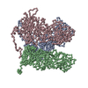
| |||||||||||||||||||||||||||||||||||||||||||||||||||||||||||||||||||||||||||||||||||||||||||||||
| Noncrystallographic symmetry (NCS) | NCS domain:
NCS domain segments: Component-ID: _ / Refine code: _
NCS ensembles :
|
-Components
| #1: Protein | Mass: 58402.820 Da / Num. of mol.: 3 Source method: isolated from a genetically manipulated source Source: (gene. exp.) Calicivirus isolate 2117 / Production host:  Trichoplusia ni (cabbage looper) / References: UniProt: Q6VCZ3 Trichoplusia ni (cabbage looper) / References: UniProt: Q6VCZ3 |
|---|
-Experimental details
-Experiment
| Experiment | Method: ELECTRON MICROSCOPY |
|---|---|
| EM experiment | Aggregation state: PARTICLE / 3D reconstruction method: single particle reconstruction |
- Sample preparation
| Component | Name: Calicivirus isolate 2117 / Type: VIRUS Details: The major capsid protein VP1 was expressed in a baculovirus system - in Hi5 cells Entity ID: all / Source: RECOMBINANT |
|---|---|
| Molecular weight | Experimental value: NO |
| Source (natural) | Organism: Calicivirus isolate 2117 |
| Source (recombinant) | Organism: Trichoplusia ni (cabbage looper) |
| Details of virus | Empty: YES / Enveloped: NO / Isolate: STRAIN / Type: VIRUS-LIKE PARTICLE |
| Natural host | Organism: unidentified |
| Virus shell | Name: VP1 / Diameter: 400 nm / Triangulation number (T number): 3 |
| Buffer solution | pH: 7.4 / Details: Phosphate buffered saline |
| Specimen | Conc.: 0.2 mg/ml / Embedding applied: NO / Shadowing applied: NO / Staining applied: NO / Vitrification applied: YES / Details: Virus-like particles |
| Specimen support | Grid material: COPPER / Grid mesh size: 400 divisions/in. / Grid type: C-flat-1.2/1.3 |
| Vitrification | Instrument: FEI VITROBOT MARK IV / Cryogen name: ETHANE / Humidity: 95 % / Chamber temperature: 277 K Details: Sample was loaded onto C-flat C1.2/1.3 grids bearing a thin continuous carbon film, allowed to adsorb for one minute before blotting for 3 seconds and plunging into liquid ethane. |
- Electron microscopy imaging
| Microscopy | Model: JEOL CRYO ARM 300 |
|---|---|
| Electron gun | Electron source:  FIELD EMISSION GUN / Accelerating voltage: 300 kV / Illumination mode: FLOOD BEAM FIELD EMISSION GUN / Accelerating voltage: 300 kV / Illumination mode: FLOOD BEAM |
| Electron lens | Mode: BRIGHT FIELD / Cs: 2.7 mm / Alignment procedure: ZEMLIN TABLEAU |
| Specimen holder | Cryogen: NITROGEN / Specimen holder model: JEOL CRYOSPECPORTER |
| Image recording | Average exposure time: 2 sec. / Electron dose: 55 e/Å2 / Detector mode: INTEGRATING / Film or detector model: DIRECT ELECTRON DE-64 (8k x 8k) / Num. of grids imaged: 5 / Num. of real images: 2000 Details: Data collection was performed as part of installation of the new CryoARM 300 microscope - five short sessions were run using primarily JADAS although one collection was performed using SerialEM. |
| Image scans | Width: 8192 / Height: 8192 / Movie frames/image: 50 / Used frames/image: 1-50 |
- Processing
| Software | Name: PHENIX / Version: 1.18.2_3874: / Classification: refinement | ||||||||||||||||||||||||||||||||||||||||||||
|---|---|---|---|---|---|---|---|---|---|---|---|---|---|---|---|---|---|---|---|---|---|---|---|---|---|---|---|---|---|---|---|---|---|---|---|---|---|---|---|---|---|---|---|---|---|
| EM software |
| ||||||||||||||||||||||||||||||||||||||||||||
| CTF correction | Type: PHASE FLIPPING AND AMPLITUDE CORRECTION | ||||||||||||||||||||||||||||||||||||||||||||
| Particle selection | Num. of particles selected: 65071 Details: Automated particle picking was performed using RELION | ||||||||||||||||||||||||||||||||||||||||||||
| Symmetry | Point symmetry: I (icosahedral) | ||||||||||||||||||||||||||||||||||||||||||||
| 3D reconstruction | Resolution: 3.65 Å / Resolution method: FSC 0.143 CUT-OFF / Num. of particles: 15989 / Num. of class averages: 1 / Symmetry type: POINT | ||||||||||||||||||||||||||||||||||||||||||||
| Atomic model building | Protocol: AB INITIO MODEL | ||||||||||||||||||||||||||||||||||||||||||||
| Refinement | Resolution: 3.65→3.65 Å / Cor.coef. Fo:Fc: 0.478 / SU B: 141.154 / SU ML: 2.046 / ESU R: 0.481 Stereochemistry target values: MAXIMUM LIKELIHOOD WITH PHASES
| ||||||||||||||||||||||||||||||||||||||||||||
| Solvent computation | Solvent model: PARAMETERS FOR MASK CACLULATION | ||||||||||||||||||||||||||||||||||||||||||||
| Displacement parameters | Biso mean: 97.957 Å2
| ||||||||||||||||||||||||||||||||||||||||||||
| Refinement step | Cycle: 1 / Total: 11902 | ||||||||||||||||||||||||||||||||||||||||||||
| Refine LS restraints |
| ||||||||||||||||||||||||||||||||||||||||||||
| Refine LS restraints NCS | Refine-ID: ELECTRON MICROSCOPY / Type: interatomic distance / Weight position: 0.05
| ||||||||||||||||||||||||||||||||||||||||||||
| LS refinement shell | Resolution: 3.8→3.899 Å / Total num. of bins used: 20
|
