 Movie
Movie Controller


+ Open data

- Basic information
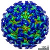
| Entry | Database: PDB / ID: 6p4j | ||||||
|---|---|---|---|---|---|---|---|
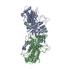
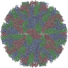
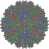
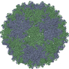
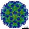
| Title | Mouse norovirus complexed with GCDCA | ||||||
 Components Components | Capsid protein | ||||||
 Keywords Keywords | VIRUS / norovirus / bile salts | ||||||
| Function / homology |  Function and homology information Function and homology informationantigenic variation / symbiont-mediated perturbation of host defense response / virion component / host cell cytoplasm Similarity search - Function | ||||||
| Biological species |  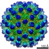 Murine norovirus 1 Murine norovirus 1 | ||||||
| Method | ELECTRON MICROSCOPY / single particle reconstruction / cryo EM / Resolution: 3.1 Å | ||||||
 Authors Authors | Smith, T.J. / Smith, T.J. | ||||||
| Funding support |  United States, 1items United States, 1items
| ||||||
 Citation Citation | Journal: J Virol / Year: 2019 Title: Bile Salts Alter the Mouse Norovirus Capsid Conformation: Possible Implications for Cell Attachment and Immune Evasion. Authors: Michael B Sherman / Alexis N Williams / Hong Q Smith / Christopher Nelson / Craig B Wilen / Daved H Fremont / Herbert W Virgin / Thomas J Smith / Abstract: Caliciviruses are single-stranded RNA viruses with 180 copies of capsid protein comprising the T=3 icosahedral capsids. The main capsid feature is a pronounced protruding (P) domain dimer formed by ...Caliciviruses are single-stranded RNA viruses with 180 copies of capsid protein comprising the T=3 icosahedral capsids. The main capsid feature is a pronounced protruding (P) domain dimer formed by adjacent subunits on the icosahedral surface while the shell domain forms a tight icosahedral sphere around the genome. While the P domain in the crystal structure of human Norwalk virus (genotype I.1) was tightly associated with the shell surface, the cryo-electron microscopy (cryo-EM) structures of several members of the family (mouse norovirus [MNV], rabbit hemorrhagic disease virus, and human norovirus genotype II.10) revealed a "floating" P domain that hovers above the shell by nearly 10 to 15 Å in physiological buffers. Since this unusual feature is shared among, and unique to, the , it suggests an important biological role. Recently, we demonstrated that bile salts enhance cell attachment to the target cell and increase the intrinsic affinity between the P domain and receptor. Presented here are the cryo-EM structures of MNV-1 in the presence of bile salts (∼3 Å) and the receptor CD300lf (∼8 Å). Surprisingly, bile salts cause the rotation and contraction of the P domain onto the shell surface. This both stabilizes the P domain and appears to allow for a higher degree of saturation of receptor onto the virus. Together, these results suggest that, as the virus moves into the gut and the associated high concentrations of bile, the entire capsid face undergoes a conformational change to optimize receptor avidity while the P domain itself undergoes smaller conformational changes to improve receptor affinity. Mouse norovirus and several other members of the have been shown to have a highly unusual structure with the receptor binding protruding (P) domain only loosely tethered to the main capsid shell. Recent studies demonstrated that bile salts enhance the intrinsic P domain/receptor affinity and is necessary for cell attachment. Presented here are the high-resolution cryo-EM structures of apo MNV, MNV/bile salt, and MNV/bile salt/receptor. Bile salts cause a 90° rotation and collapse of the P domain onto the shell surface that may increase the number of available receptor binding sites. Therefore, bile salts appear to be having several effects on MNV. Bile salts shift the structural equilibrium of the P domain toward a form that binds the receptor and away from one that binds antibody. They may also cause the entire P domain to optimize receptor binding while burying a number of potential epitopes. | ||||||
| History |
|
- Structure visualization
| Movie |
Movie viewer |
|---|---|
| Structure viewer | Molecule: MolmilJmol/JSmol |
- Downloads & links
-Download
| PDBx/mmCIF format | 6p4j.cif.gz | 272 KB | Display | PDBx/mmCIF format |
|---|---|---|---|---|
| PDB format | pdb6p4j.ent.gz | 223 KB | Display | PDB format |
| PDBx/mmJSON format | 6p4j.json.gz | Tree view | PDBx/mmJSON format | |
| Others |  Other downloads Other downloads |
-Validation report
| Arichive directory | https://data.pdbj.org/pub/pdb/validation_reports/p4/6p4jftp://data.pdbj.org/pub/pdb/validation_reports/p4/6p4j | HTTPS FTP |
|---|
-Related structure data
| Related structure data | 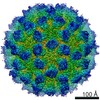 20250MC  6p4kC  6p4lC M: map data used to model this data C: citing same article ( |
|---|---|
| Similar structure data |


-Links
 PDBj
PDBj
- Assembly
| Deposited unit | 
|
|---|---|
| 1 | x 60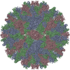
|
| 2 |
|
| 3 | x 5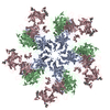
|
| 4 | x 6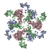
|
| 5 | 
|
| Symmetry | Point symmetry: (Schoenflies symbol: I (icosahedral)) |
-Components
| #1: Protein | Mass: 56122.590 Da / Num. of mol.: 3 / Source method: isolated from a natural source / Source: (natural) Murine norovirus 1 / References: UniProt: Q2V8W4#2: Chemical | ChemComp-CHO / 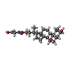  Mass: 449.623 Da / Num. of mol.: 6 / Source method: obtained synthetically / Formula: C26H43NO5 / Comment: detergent*YM Mass: 449.623 Da / Num. of mol.: 6 / Source method: obtained synthetically / Formula: C26H43NO5 / Comment: detergent*YM |
|---|
-Experimental details
-Experiment
| Experiment | Method: ELECTRON MICROSCOPY |
|---|---|
| EM experiment | Aggregation state: PARTICLE / 3D reconstruction method: single particle reconstruction |
- Sample preparation
| Component | Name: Murine adenovirus 1 / Type: VIRUS / Entity ID: #1 / Source: NATURAL |
|---|---|
| Source (natural) | Organism: Murine adenovirus 1 |
| Details of virus | Empty: NO / Enveloped: NO / Isolate: SPECIES / Type: VIRION |
| Natural host | Organism: Mus musculus |
| Buffer solution | pH: 7.2 |
| Specimen | Embedding applied: NO / Shadowing applied: NO / Staining applied: NO / Vitrification applied: YES |
| Vitrification | Cryogen name: ETHANE |
- Electron microscopy imaging
| Experimental equipment |  Model: Titan Krios / Image courtesy: FEI Company |
|---|---|
| Microscopy | Model: FEI TITAN KRIOS |
| Electron gun | Electron source:  FIELD EMISSION GUN / Accelerating voltage: 300 kV / Illumination mode: SPOT SCAN FIELD EMISSION GUN / Accelerating voltage: 300 kV / Illumination mode: SPOT SCAN |
| Electron lens | Mode: BRIGHT FIELD |
| Image recording | Electron dose: 38 e/Å2 / Film or detector model: OTHER |
- Processing
| CTF correction | Type: PHASE FLIPPING AND AMPLITUDE CORRECTION |
|---|---|
| 3D reconstruction | Resolution: 3.1 Å / Resolution method: FSC 0.143 CUT-OFF / Num. of particles: 28463 / Symmetry type: POINT |
