 Movie
Movie Controller

[English] 日本語
 Yorodumi
- EMDB-12194: The cryo-EM structure of vesivirus 2117, an adventitious agent an... -
+ Open data

- Basic information
| Entry | Database: EMDB / ID: EMD-12194 | |||||||||
|---|---|---|---|---|---|---|---|---|---|---|
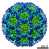
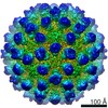
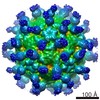
| Title | The cryo-EM structure of vesivirus 2117, an adventitious agent and possible cause of haemorrhagic gastroenteritis in dogs. | |||||||||
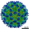
 Map data Map data | Vesivirus 2117 virus-like particle - sharpened map | |||||||||
 Sample Sample |
| |||||||||
 Keywords Keywords | capsid / calicivirus / vp1 / VIRAL PROTEIN | |||||||||
| Function / homology | T=3 icosahedral viral capsid / Calicivirus coat protein / Calicivirus coat protein / Picornavirus/Calicivirus coat protein / Viral coat protein subunit / host cell cytoplasm / Capsid protein VP1 Function and homology information Function and homology information | |||||||||
| Biological species |  Calicivirus isolate 2117 Calicivirus isolate 2117 | |||||||||
| Method | single particle reconstruction / cryo EM / Resolution: 3.65 Å | |||||||||
 Authors Authors | Sutherland H / Conley MJ | |||||||||
| Funding support |  United Kingdom, 2 items United Kingdom, 2 items
| |||||||||
 Citation Citation | Journal: J Virol / Year: 2021 Title: The Cryo-EM Structure of Vesivirus 2117 Highlights Functional Variations in Entry Pathways for Viruses in Different Clades of the Genus. Authors: Hazel Sutherland / Michaela J Conley / Edward Emmott / James Streetley / Ian G Goodfellow / David Bhella / Abstract: Vesivirus 2117 is an adventitious agent that has been responsible for lost productivity in biopharmaceutical production following contamination of Chinese hamster ovary cell cultures in commercial ...Vesivirus 2117 is an adventitious agent that has been responsible for lost productivity in biopharmaceutical production following contamination of Chinese hamster ovary cell cultures in commercial bioreactors. A member of the , 2117 is classified within the Vesivirus genus in a clade that includes canine and mink caliciviruses but is distinct from the vesicular exanthema of swine virus (VESV) clade, which includes the extensively studied feline calicivirus (FCV). We have used cryogenic electron microscopy (cryo-EM) to determine the structure of the capsid of this small, icosahedral, positive-sense-RNA-containing virus. We show that the outer face of the dimeric capsomeres, which contains the receptor binding site and major immunodominant epitopes in all caliciviruses studied thus far, is quite different from that of FCV. This is a consequence of a 22-amino-acid insertion in the sequence of the FCV major capsid protein that forms a "cantilevered arm" that both plays an important role in receptor engagement and undergoes structural rearrangements thought to be important for genome delivery to the cytosol. Our data highlight a potentially important difference in the attachment and entry pathways employed by the different clades of the genus. Vesivirus 2117 has caused significant losses in manufacturing of biopharmaceutical products following contamination of cell cultures used in their production. We report the structure of the vesivirus 2117 capsid, the shell that encloses the virus's genome. Comparison of this structure with that of a related vesivirus, feline calicivirus (FCV), highlighted potentially important differences related to virus attachment and entry. Our findings suggest that these two viruses may bind differently to receptors at the host cell surface. We also show that a region of the capsid protein of FCV that rearranges following receptor engagement is not present in vesivirus 2117. These structural changes in the FCV capsid have been shown to allow the assembly of a portal-like structure that is hypothesized to deliver the viral genome to the cell's interior. Our data suggest that the 2117 portal assembly may employ a different means of anchoring to the outer face of the capsid. #1: Journal: Biorxiv / Year: 2021Title: The cryo-EM structure of vesivirus 2117 highlights functional variations in entry pathways for viruses in different clades of the Vesivirus genus Authors: Sutherland H / Conley MJ / Emmott E / Streetley J / Goodfellow IG / Bhella D | |||||||||
| History |
|
- Structure visualization
| Movie |
Movie viewer |
|---|---|
| Structure viewer | EM map: SurfViewMolmilJmol/JSmol |
| Supplemental images |

- Downloads & links
-EMDB archive
| Map data | emd_12194.map.gz | 468.6 MB | EMDB map data format | |
|---|---|---|---|---|
| Header (meta data) | emd-12194-v30.xmlemd-12194.xml | 17.9 KB 17.9 KB | Display Display | EMDB header |
| FSC (resolution estimation) | emd_12194_fsc.xml | 18.1 KB | Display | FSC data file |
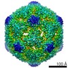
| Images |  emd_12194.png emd_12194.png | 237.1 KB | ||
| Filedesc metadata | emd-12194.cif.gz | 6.5 KB | ||
| Others | emd_12194_additional_1.map.gz | 474.1 MB | ||
| Archive directory |  http://ftp.pdbj.org/pub/emdb/structures/EMD-12194ftp://ftp.pdbj.org/pub/emdb/structures/EMD-12194 http://ftp.pdbj.org/pub/emdb/structures/EMD-12194ftp://ftp.pdbj.org/pub/emdb/structures/EMD-12194 | HTTPS FTP |
-Related structure data
| Related structure data |  7bjpMC M: atomic model generated by this map C: citing same article ( |
|---|---|
| Similar structure data | |
| EM raw data | EMPIAR-10638 (Title: The cryo-EM structure of vesivirus 2117 highlights functional variations in entry pathways for viruses in different clades of the vesivirus genus. Data size: 24.4 TB Data #1: Unaligned movies of calicivirus 2117 VLPs [micrographs - multiframe]) |


-Links
| EMDB pages | EMDB (EBI/PDBe) / EMDataResource |
|---|---|
| Related items in Molecule of the Month |


-Map
| File | Download / File: emd_12194.map.gz / Format: CCP4 / Size: 512 MB / Type: IMAGE STORED AS FLOATING POINT NUMBER (4 BYTES) | ||||||||||||||||||||||||||||||||||||||||||||||||||||||||||||
|---|---|---|---|---|---|---|---|---|---|---|---|---|---|---|---|---|---|---|---|---|---|---|---|---|---|---|---|---|---|---|---|---|---|---|---|---|---|---|---|---|---|---|---|---|---|---|---|---|---|---|---|---|---|---|---|---|---|---|---|---|---|
| Annotation | Vesivirus 2117 virus-like particle - sharpened map | ||||||||||||||||||||||||||||||||||||||||||||||||||||||||||||
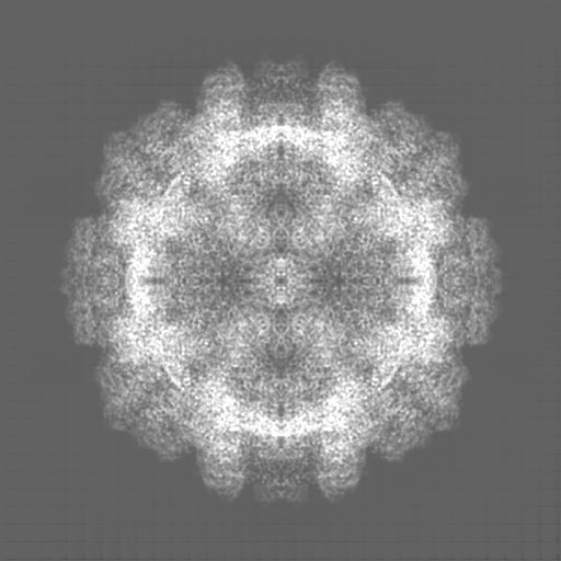
| Projections & slices | Image control
Images are generated by Spider. | ||||||||||||||||||||||||||||||||||||||||||||||||||||||||||||
| Voxel size | X=Y=Z: 0.998 Å | ||||||||||||||||||||||||||||||||||||||||||||||||||||||||||||
| Density |
| ||||||||||||||||||||||||||||||||||||||||||||||||||||||||||||
| Symmetry | Space group: 1 | ||||||||||||||||||||||||||||||||||||||||||||||||||||||||||||
| Details | EMDB XML:
CCP4 map header:
| ||||||||||||||||||||||||||||||||||||||||||||||||||||||||||||
 Z (Sec.)
 Y (Row.)
 X (Col.)


-Supplemental data
-Additional map: Vesivirus 2117 virus-like particle - DeepEMhancer modified map
| File | emd_12194_additional_1.map | ||||||||||||
|---|---|---|---|---|---|---|---|---|---|---|---|---|---|
| Annotation | Vesivirus 2117 virus-like particle - DeepEMhancer modified map | ||||||||||||
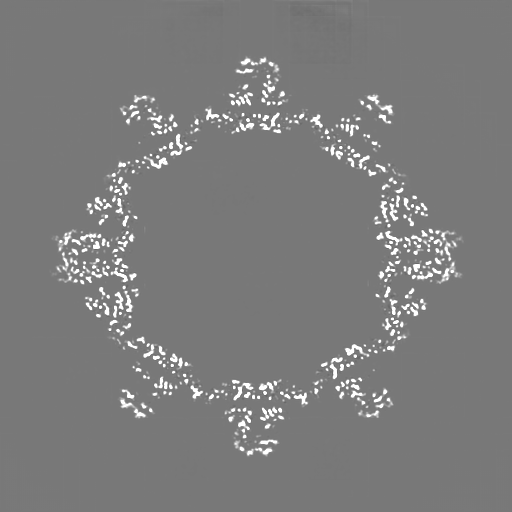
| Projections & Slices |
| ||||||||||||
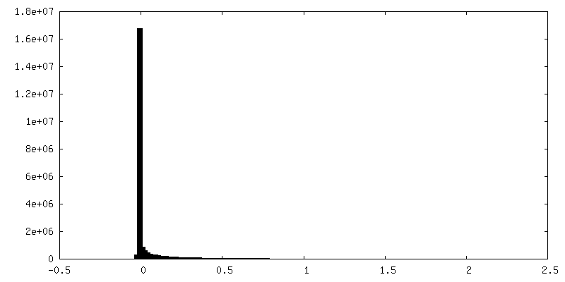
| Density Histograms |


- Sample components
-Entire : Calicivirus isolate 2117
| Entire | Name: Calicivirus isolate 2117 |
|---|---|
| Components |
|
-Supramolecule #1: Calicivirus isolate 2117
| Supramolecule | Name: Calicivirus isolate 2117 / type: virus / ID: 1 / Parent: 0 / Macromolecule list: all Details: The major capsid protein VP1 was expressed in a baculovirus system - in Hi5 cells NCBI-ID: 241631 / Sci species name: Calicivirus isolate 2117 / Virus type: VIRUS-LIKE PARTICLE / Virus isolate: STRAIN / Virus enveloped: No / Virus empty: Yes |
|---|---|
| Host (natural) | Organism: unidentified (others) |
| Virus shell | Shell ID: 1 / Name: VP1 / Diameter: 400.0 Å / T number (triangulation number): 3 |
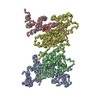
-Macromolecule #1: Capsid protein
| Macromolecule | Name: Capsid protein / type: protein_or_peptide / ID: 1 / Number of copies: 3 / Enantiomer: LEVO |
|---|---|
| Source (natural) | Organism: Calicivirus isolate 2117 |
| Molecular weight | Theoretical: 58.40282 KDa |
| Recombinant expression | Organism:  Trichoplusia ni (cabbage looper) Trichoplusia ni (cabbage looper) |
| Sequence | String: MSGDASSGSP DITGEEQGVA VATGTQPSAP AMATLATAAT GTMPEEWKTF FSYYTTVNWS TTDETGKVLF VQGLSPRMNP FLDHLAKMY TGWSGSMEIR FTISGSGVFG GKLAAVMVPP GIGTEGGTSL LQFPHVLVDA RQTEPVIFNI PDIRTVLWHD M HDTLTAHL ...String: MSGDASSGSP DITGEEQGVA VATGTQPSAP AMATLATAAT GTMPEEWKTF FSYYTTVNWS TTDETGKVLF VQGLSPRMNP FLDHLAKMY TGWSGSMEIR FTISGSGVFG GKLAAVMVPP GIGTEGGTSL LQFPHVLVDA RQTEPVIFNI PDIRTVLWHD M HDTLTAHL VILVYNDLLN PYQNTTTGTS CTVTVETRGG TDFEFHLLKP PSRKMIFGAD PSRLIPKKSM FWEGNRLPGE FK GFSIKPL VFQANRHFDC KRQTFGWSTP EHAGVKLNIQ RQNLDTEDKT DIGVHLVTGL KTIKSQVPDG WPDYYGRNII LAN TTASFG EVSEAMLGTV VPYRVSGKLE WRHLPEIAFA NGTAKNSTIV CGKYLTGNFY VGGNFTQQGN VVVYPAFWTS KHGD TKCIG EDEDMVKRID VLPQAQTTGG NYPIYYVTEF PAAYLPAPRV YNSQLLWTSR LLAQDVYDIG PEALAVFKIK DSAGN WFDI GISCEGFSFV GAPTLPFSSL QFPLEASYVG MASAYNKLQH NIAGTSVTL UniProtKB: Capsid protein VP1 |
-Experimental details
-Structure determination
| Method | cryo EM |
|---|---|
 Processing Processing | single particle reconstruction |
| Aggregation state | particle |
-Sample preparation
| Concentration | 0.2 mg/mL |
|---|---|
| Buffer | pH: 7.4 / Details: Phosphate buffered saline |
| Grid | Model: C-flat-1.2/1.3 / Material: COPPER / Mesh: 400 / Support film - Material: CARBON / Support film - topology: CONTINUOUS / Pretreatment - Type: GLOW DISCHARGE |
| Vitrification | Cryogen name: ETHANE / Chamber humidity: 95 % / Chamber temperature: 277 K / Instrument: FEI VITROBOT MARK IV Details: Sample was loaded onto C-flat C1.2/1.3 grids bearing a thin continuous carbon film, allowed to adsorb for one minute before blotting for 3 seconds and plunging into liquid ethane.. |
| Details | Virus-like particles |
- Electron microscopy
| Microscope | JEOL CRYO ARM 300 |
|---|---|
| Image recording | Film or detector model: DIRECT ELECTRON DE-64 (8k x 8k) / Detector mode: INTEGRATING / Digitization - Dimensions - Width: 8192 pixel / Digitization - Dimensions - Height: 8192 pixel / Digitization - Frames/image: 1-50 / Number grids imaged: 5 / Number real images: 2000 / Average exposure time: 2.0 sec. / Average electron dose: 55.0 e/Å2 Details: Data collection was performed as part of installation of the new CryoARM 300 microscope - five short sessions were run using primarily JADAS although one collection was performed using SerialEM. |
| Electron beam | Acceleration voltage: 300 kV / Electron source:  FIELD EMISSION GUN FIELD EMISSION GUN |
| Electron optics | Illumination mode: FLOOD BEAM / Imaging mode: BRIGHT FIELD / Cs: 2.7 mm |
| Sample stage | Specimen holder model: JEOL CRYOSPECPORTER / Cooling holder cryogen: NITROGEN |
+Image processing


-Atomic model buiding 1
| Refinement | Protocol: AB INITIO MODEL |
|---|---|
| Output model | PDB-7bjp: |
