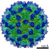
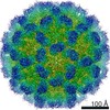
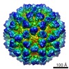
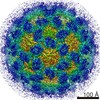
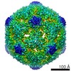
Journal: J Virol / Year: 2015 Title: Antigenic and Cryo-Electron Microscopy Structure Analysis of a Chimeric Sapovirus Capsid. Authors: Naoyuki Miyazaki / David W Taylor / Grant S Hansman / Kazuyoshi Murata / Abstract: The capsid protein (VP1) of all caliciviruses forms an icosahedral particle with two principal domains, shell (S) and protruding (P) domains, which are connected via a flexible hinge region. The S ...The capsid protein (VP1) of all caliciviruses forms an icosahedral particle with two principal domains, shell (S) and protruding (P) domains, which are connected via a flexible hinge region. The S domain forms a scaffold surrounding the nucleic acid, while the P domains form a homodimer that interacts with receptors. The P domain is further subdivided into two subdomains, termed P1 and P2. The P2 subdomain is likely an insertion in the P1 subdomain; consequently, the P domain is divided into the P1-1, P2, and P1-2 subdomains. In order to investigate capsid antigenicity, N-terminal (N-term)/S/P1-1 and P2/P1-2 were switched between two sapovirus genotypes GI.1 and GI.5. The chimeric VP1 constructs were expressed in insect cells and were shown to self-assemble into virus-like particles (VLPs) morphologically similar to the parental VLPs. Interestingly, the chimeric VLPs had higher levels of cross-reactivities to heterogeneous antisera than the parental VLPs. In order to better understand the antigenicity from a structural perspective, we determined an intermediate-resolution (8.5-Å) cryo-electron microscopy (cryo-EM) structure of a chimeric VLP and developed a VP1 homology model. The cryo-EM structure revealed that the P domain dimers were raised slightly (∼5 Å) above the S domain. The VP1 homology model allowed us predict the S domain (67-229) and P1-1 (229-280), P2 (281-447), and P1-2 (448-567) subdomains. Our results suggested that the raised P dimers might expose immunoreactive S/P1-1 subdomain epitopes. Consequently, the higher levels of cross-reactivities with the chimeric VLPs resulted from a combination of GI.1 and GI.5 epitopes. IMPORTANCE: We developed sapovirus chimeric VP1 constructs and produced the chimeric VLPs in insect cells. We found that both chimeric VLPs had a higher level of cross-reactivity against ...IMPORTANCE: We developed sapovirus chimeric VP1 constructs and produced the chimeric VLPs in insect cells. We found that both chimeric VLPs had a higher level of cross-reactivity against heterogeneous VLP antisera than the parental VLPs. The cryo-EM structure of one chimeric VLP (Yokote/Mc114) was solved to 8.5-Å resolution. A homology model of the VP1 indicated for the first time the putative S and P (P1-1, P2, and P1-2) domains. The overall structure of Yokote/Mc114 contained features common among other caliciviruses. We showed that the P2 subdomain was mainly involved in the homodimeric interface, whereas a large gap between the P1 subdomains had fewer interactions.
History
Deposition
Dec 14, 2015
-
Header (metadata) release
Jan 27, 2016
-
Map release
Feb 10, 2016
-
Update
Feb 8, 2017
-
Current status
Feb 8, 2017
Processing site: PDBe / Status: Released
-
Structure visualization
Movie
Surface view with section colored by density value
Supramolecule #1000: Chimera human sapovirus capsid
Supramolecule
Name: Chimera human sapovirus capsid / type: sample / ID: 1000 / Oligomeric state: icosahedral / Number unique components: 1
Molecular weight
Experimental: 10 MDa / Theoretical: 10 MDa
-
Supramolecule #1: Sapovirus
Supramolecule
Name: Sapovirus / type: virus / ID: 1 / Name.synonym: human sapovirus / NCBI-ID: 95341 / Sci species name: Sapovirus / Virus type: VIRUS-LIKE PARTICLE / Virus isolate: OTHER / Virus enveloped: No / Virus empty: Yes / Syn species name: human sapovirus
Host (natural)
Organism: Homo sapiens (human) / synonym: VERTEBRATES
Host system
Organism: unidentified baculovirus
Molecular weight
Experimental: 10 MDa / Theoretical: 10 MDa
Virus shell
Shell ID: 1 / Diameter: 38 Å / T number (triangulation number): 3
-
Experimental details
-
Structure determination
Method
negative staining, cryo EM
Processing
single particle reconstruction
Aggregation state
particle
-
Sample preparation
Staining
Type: NEGATIVE / Details: ice-embedding
Grid
Details: quantifoil R1.2/1.3
Vitrification
Cryogen name: ETHANE / Chamber humidity: 95 % / Chamber temperature: 120 K / Instrument: FEI VITROBOT MARK III
-
Electron microscopy
Microscope
JEOL 2200FS
Temperature
Min: 90 K / Max: 105 K / Average: 100 K
Specialist optics
Energy filter - Name: Omega-type / Energy filter - Lower energy threshold: 0.0 eV / Energy filter - Upper energy threshold: 15.0 eV
Date
Jul 15, 2012
Image recording
Category: CCD / Film or detector model: TVIPS TEMCAM-F415 (4k x 4k) / Digitization - Sampling interval: 15 µm / Number real images: 80 / Average electron dose: 20 e/Å2 / Od range: 5 / Bits/pixel: 16
Electron beam
Acceleration voltage: 200 kV / Electron source: FIELD EMISSION GUN
Applied symmetry - Point group: I (icosahedral) / Algorithm: OTHER / Resolution.type: BY AUTHOR / Resolution: 8.5 Å / Resolution method: OTHER / Software - Name: EMAN2 / Number images used: 6000
+
About Yorodumi
-
News
-
Feb 9, 2022. New format data for meta-information of EMDB entries
New format data for meta-information of EMDB entries
Version 3 of the EMDB header file is now the official format.
The previous official version 1.9 will be removed from the archive.
In the structure databanks used in Yorodumi, some data are registered as the other names, "COVID-19 virus" and "2019-nCoV". Here are the details of the virus and the list of structure data.
Jan 31, 2019. EMDB accession codes are about to change! (news from PDBe EMDB page)
EMDB accession codes are about to change! (news from PDBe EMDB page)
The allocation of 4 digits for EMDB accession codes will soon come to an end. Whilst these codes will remain in use, new EMDB accession codes will include an additional digit and will expand incrementally as the available range of codes is exhausted. The current 4-digit format prefixed with “EMD-” (i.e. EMD-XXXX) will advance to a 5-digit format (i.e. EMD-XXXXX), and so on. It is currently estimated that the 4-digit codes will be depleted around Spring 2019, at which point the 5-digit format will come into force.
The EM Navigator/Yorodumi systems omit the EMD- prefix.
Related info.:Q: What is EMD? / ID/Accession-code notation in Yorodumi/EM Navigator
Yorodumi is a browser for structure data from EMDB, PDB, SASBDB, etc.
This page is also the successor to EM Navigator detail page, and also detail information page/front-end page for Omokage search.
The word "yorodu" (or yorozu) is an old Japanese word meaning "ten thousand". "mi" (miru) is to see.
Related info.:EMDB / PDB / SASBDB / Comparison of 3 databanks / Yorodumi Search / Aug 31, 2016. New EM Navigator & Yorodumi / Yorodumi Papers / Jmol/JSmol / Function and homology information / Changes in new EM Navigator and Yorodumi
 Movie
Movie Controller

 Yorodumi
 Open data
 Basic information
 Map data
 Sample
 Keywords
 Sapovirus
 Authors
 Citation


 Structure visualization
 Movie viewer
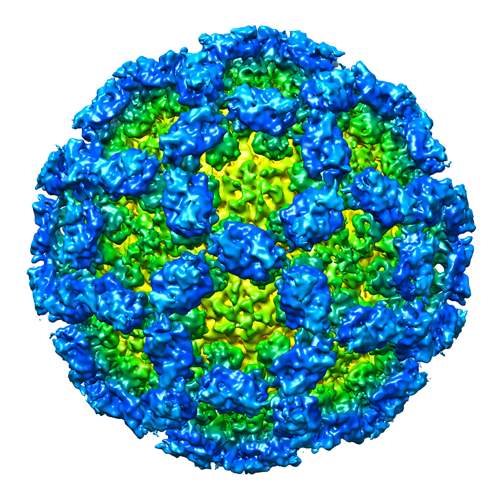
 Downloads & links
 EMD-3281.png
 http://ftp.pdbj.org/pub/emdb/structures/EMD-3281
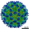
 Z (Sec.)
 Y (Row.)
 X (Col.)


 Sample components
 Homo sapiens (human) / synonym: VERTEBRATES
 Processing
 Electron microscopy
 FIELD EMISSION GUN