 Movie
Movie Controller

[English] 日本語
 Yorodumi
- EMDB-5410: Cryo-electron microscopic reconstruction of wild rabbit hemorrhag... -
+ Open data

- Basic information
| Entry | Database: EMDB / ID: EMD-5410 | |||||||||
|---|---|---|---|---|---|---|---|---|---|---|
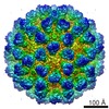
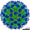
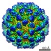
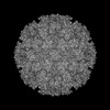
| Title | Cryo-electron microscopic reconstruction of wild rabbit hemorrhagic disease virus | |||||||||
 Map data Map data | Reconstruction of wild rabbit hemorrhagic disease virus; The final map was sharpened using b factor -300 A2 to 4.5 angstrom and low passed to 5 angstrom. | |||||||||
 Sample Sample |
| |||||||||
 Keywords Keywords | calicivirus / Lagovirus / rabbit hemorrhagic disease | |||||||||
| Function / homology |  Function and homology information Function and homology informationviral capsid / ribonucleoside triphosphate phosphatase activity / host cell cytoplasm / RNA helicase activity / cysteine-type endopeptidase activity / viral RNA genome replication / RNA-directed RNA polymerase activity / DNA-templated transcription / proteolysis / RNA binding / ATP binding Similarity search - Function | |||||||||
| Biological species |  Rabbit hemorrhagic disease virus Rabbit hemorrhagic disease virus | |||||||||
| Method | single particle reconstruction / cryo EM / Resolution: 6.5 Å | |||||||||
 Authors Authors | Wang X / Xu F / Liu J / Gao B / Zhang K / Zhai Y / Ma J / Hu Z / Pang X / Zheng D ...Wang X / Xu F / Liu J / Gao B / Zhang K / Zhai Y / Ma J / Hu Z / Pang X / Zheng D / Pang H / Sun F | |||||||||
 Citation Citation | Journal: PLoS Pathog / Year: 2013 Title: Atomic model of rabbit hemorrhagic disease virus by cryo-electron microscopy and crystallography. Authors: Xue Wang / Fengting Xu / Jiasen Liu / Bingquan Gao / Yanxin Liu / Yujia Zhai / Jun Ma / Kai Zhang / Timothy S Baker / Klaus Schulten / Dong Zheng / Hai Pang / Fei Sun /  Abstract: Rabbit hemorrhagic disease, first described in China in 1984, causes hemorrhagic necrosis of the liver. Its etiological agent, rabbit hemorrhagic disease virus (RHDV), belongs to the Lagovirus genus ...Rabbit hemorrhagic disease, first described in China in 1984, causes hemorrhagic necrosis of the liver. Its etiological agent, rabbit hemorrhagic disease virus (RHDV), belongs to the Lagovirus genus in the family Caliciviridae. The detailed molecular structure of any lagovirus capsid has yet to be determined. Here, we report a cryo-electron microscopic (cryoEM) reconstruction of wild-type RHDV at 6.5 Å resolution and the crystal structures of the shell (S) and protruding (P) domains of its major capsid protein, VP60, each at 2.0 Å resolution. From these data we built a complete atomic model of the RHDV capsid. VP60 has a conserved S domain and a specific P2 sub-domain that differs from those found in other caliciviruses. As seen in the shell portion of the RHDV cryoEM map, which was resolved to ~5.5 Å, the N-terminal arm domain of VP60 folds back onto its cognate S domain. Sequence alignments of VP60 from six groups of RHDV isolates revealed seven regions of high variation that could be mapped onto the surface of the P2 sub-domain and suggested three putative pockets might be responsible for binding to histo-blood group antigens. A flexible loop in one of these regions was shown to interact with rabbit tissue cells and contains an important epitope for anti-RHDV antibody production. Our study provides a reliable, pseudo-atomic model of a Lagovirus and suggests a new candidate for an efficient vaccine that can be used to protect rabbits from RHDV infection. | |||||||||
| History |
|
- Structure visualization
| Movie |
Movie viewer |
|---|---|
| Structure viewer | EM map: SurfViewMolmilJmol/JSmol |
| Supplemental images |

- Downloads & links
-EMDB archive
| Map data | emd_5410.map.gz | 326.3 MB | EMDB map data format | |
|---|---|---|---|---|
| Header (meta data) | emd-5410-v30.xmlemd-5410.xml | 13.1 KB 13.1 KB | Display Display | EMDB header |
| Images | 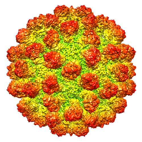 emd_5410.jpg emd_5410.jpg | 390 KB | ||
| Archive directory |  http://ftp.pdbj.org/pub/emdb/structures/EMD-5410ftp://ftp.pdbj.org/pub/emdb/structures/EMD-5410 http://ftp.pdbj.org/pub/emdb/structures/EMD-5410ftp://ftp.pdbj.org/pub/emdb/structures/EMD-5410 | HTTPS FTP |
-Related structure data
| Related structure data |  3j1pMC  4egtC  4ejrC M: atomic model generated by this map C: citing same article ( |
|---|---|
| Similar structure data |

-Links
| EMDB pages | EMDB (EBI/PDBe) / EMDataResource |
|---|---|
| Related items in Molecule of the Month |


-Map
| File | Download / File: emd_5410.map.gz / Format: CCP4 / Size: 412 MB / Type: IMAGE STORED AS FLOATING POINT NUMBER (4 BYTES) | ||||||||||||||||||||||||||||||||||||||||||||||||||||||||||||||||||||
|---|---|---|---|---|---|---|---|---|---|---|---|---|---|---|---|---|---|---|---|---|---|---|---|---|---|---|---|---|---|---|---|---|---|---|---|---|---|---|---|---|---|---|---|---|---|---|---|---|---|---|---|---|---|---|---|---|---|---|---|---|---|---|---|---|---|---|---|---|---|
| Annotation | Reconstruction of wild rabbit hemorrhagic disease virus; The final map was sharpened using b factor -300 A2 to 4.5 angstrom and low passed to 5 angstrom. | ||||||||||||||||||||||||||||||||||||||||||||||||||||||||||||||||||||
| Projections & slices | Image control
Images are generated by Spider. | ||||||||||||||||||||||||||||||||||||||||||||||||||||||||||||||||||||
| Voxel size | X=Y=Z: 0.933 Å | ||||||||||||||||||||||||||||||||||||||||||||||||||||||||||||||||||||
| Density |
| ||||||||||||||||||||||||||||||||||||||||||||||||||||||||||||||||||||
| Symmetry | Space group: 1 | ||||||||||||||||||||||||||||||||||||||||||||||||||||||||||||||||||||
| Details | EMDB XML:
CCP4 map header:
| ||||||||||||||||||||||||||||||||||||||||||||||||||||||||||||||||||||
 Z (Sec.)
 Y (Row.)
 X (Col.)


-Supplemental data
- Sample components
-Entire : Wild Rabbit Hemorrhagic Disease Virus (strain HYD)
| Entire | Name: Wild Rabbit Hemorrhagic Disease Virus (strain HYD) |
|---|---|
| Components |
|
-Supramolecule #1000: Wild Rabbit Hemorrhagic Disease Virus (strain HYD)
| Supramolecule | Name: Wild Rabbit Hemorrhagic Disease Virus (strain HYD) / type: sample / ID: 1000 / Number unique components: 1 |
|---|---|
| Molecular weight | Theoretical: 10.8 MDa |
-Supramolecule #1: Rabbit hemorrhagic disease virus
| Supramolecule | Name: Rabbit hemorrhagic disease virus / type: virus / ID: 1 / Name.synonym: RHDV / Details: HYD strain and gene bank number is AEB26305. / NCBI-ID: 11976 / Sci species name: Rabbit hemorrhagic disease virus / Database: NCBI / Virus type: VIRION / Virus isolate: STRAIN / Virus enveloped: No / Virus empty: No / Syn species name: RHDV |
|---|---|
| Host (natural) | Organism:  |
| Molecular weight | Theoretical: 10.8 MDa |
| Virus shell | Shell ID: 1 / Name: VP60 / Diameter: 440 Å / T number (triangulation number): 3 |
-Experimental details
-Structure determination
| Method | cryo EM |
|---|---|
 Processing Processing | single particle reconstruction |
| Aggregation state | particle |
-Sample preparation
| Buffer | pH: 7 / Details: 50mM Tris, 50mM NaCl, 5mM EDTA |
|---|---|
| Grid | Details: 200 mesh copper grid with holey array carbon support (GiG), glow discharged |
| Vitrification | Cryogen name: ETHANE / Chamber humidity: 100 % / Chamber temperature: 100 K / Instrument: FEI VITROBOT MARK IV / Method: Blot for 3 seconds before plunging |
- Electron microscopy
| Microscope | FEI TITAN KRIOS |
|---|---|
| Temperature | Average: 85 K |
| Alignment procedure | Legacy - Astigmatism: Objective lens astigmatism was corrected at 100,000 times magnification Legacy - Electron beam tilt params: 0 |
| Date | Jan 22, 2011 |
| Image recording | Category: CCD / Film or detector model: GATAN ULTRASCAN 4000 (4k x 4k) / Digitization - Sampling interval: 15 µm / Number real images: 1100 / Average electron dose: 20 e/Å2 / Bits/pixel: 32 |
| Tilt angle min | 0 |
| Tilt angle max | 0 |
| Electron beam | Acceleration voltage: 300 kV / Electron source:  FIELD EMISSION GUN FIELD EMISSION GUN |
| Electron optics | Calibrated magnification: 160770 / Illumination mode: FLOOD BEAM / Imaging mode: BRIGHT FIELD / Cs: 2.7 mm / Nominal defocus max: 2.5 µm / Nominal defocus min: 1.5 µm / Nominal magnification: 96000 |
| Sample stage | Specimen holder: Liquid nitrogen cooled / Specimen holder model: FEI TITAN KRIOS AUTOGRID HOLDER |
| Experimental equipment |  Model: Titan Krios / Image courtesy: FEI Company |
-Image processing
| Details | The particles were selected using an automatic selection program FindEM. |
|---|---|
| CTF correction | Details: CTF correction of each whole micrograph |
| Final reconstruction | Algorithm: OTHER / Resolution.type: BY AUTHOR / Resolution: 6.5 Å / Resolution method: FSC 0.5 CUT-OFF / Software - Name: EMAN1.9, Spider Details: The final map was sharpened to 4.5 Angstrom with B factor -300 and then filtered to 5.0 Angstrom. Number images used: 26000 |
| Final two d classification | Number classes: 475 |
-Atomic model buiding 1
| Initial model | PDB ID: Chain - Chain ID: A |
|---|---|
| Software | Name: Chimera |
| Details | Protocol: Rigid body. The P domains were separately fitted into the map by automatic rigid body fitting using Chimera (FIT IN MAP). |
| Refinement | Space: REAL / Protocol: RIGID BODY FIT |
| Output model | PDB-3j1p: |
-Atomic model buiding 2
| Initial model | PDB ID: Chain - Chain ID: A |
|---|---|
| Software | Name: Chimera |
| Details | Protocol: Rigid body. The S domains were separately fitted into the map by automatic rigid body fitting using Chimera (FIT IN MAP). |
| Refinement | Space: REAL / Protocol: RIGID BODY FIT |
| Output model | PDB-3j1p: |
