 Movie
Movie Controller

[English] 日本語
 Yorodumi
- PDB-4egt: Crystal structure of major capsid protein P domain from rabbit he... -
+ Open data

- Basic information
| Entry | Database: PDB / ID: 4egt | ||||||
|---|---|---|---|---|---|---|---|
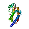
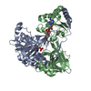
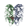
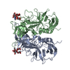
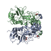
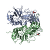
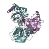
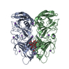
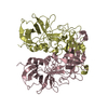
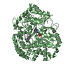
| Title | Crystal structure of major capsid protein P domain from rabbit hemorrhagic disease virus | ||||||
 Components Components | Major capsid protein VP60 | ||||||
 Keywords Keywords | VIRAL PROTEIN / capsid protein | ||||||
| Function / homology |  Function and homology information Function and homology informationvirion component / viral capsid / ribonucleoside triphosphate phosphatase activity / host cell cytoplasm / RNA helicase activity / cysteine-type endopeptidase activity / viral RNA genome replication / RNA-directed RNA polymerase activity / DNA-templated transcription / proteolysis ...virion component / viral capsid / ribonucleoside triphosphate phosphatase activity / host cell cytoplasm / RNA helicase activity / cysteine-type endopeptidase activity / viral RNA genome replication / RNA-directed RNA polymerase activity / DNA-templated transcription / proteolysis / RNA binding / ATP binding Similarity search - Function | ||||||
| Biological species |  Rabbit hemorrhagic disease virus Rabbit hemorrhagic disease virus | ||||||
| Method |  X-RAY DIFFRACTION / SYNCHROTRON / MOLECULAR REPLACEMENT / Resolution: 2 Å X-RAY DIFFRACTION / SYNCHROTRON / MOLECULAR REPLACEMENT / Resolution: 2 Å | ||||||
 Authors Authors | Wang, X. / Xu, F. / Zhang, K. / Zhai, Y. / Sun, F. | ||||||
 Citation Citation | Journal: PLoS Pathog / Year: 2013 Title: Atomic model of rabbit hemorrhagic disease virus by cryo-electron microscopy and crystallography. Authors: Xue Wang / Fengting Xu / Jiasen Liu / Bingquan Gao / Yanxin Liu / Yujia Zhai / Jun Ma / Kai Zhang / Timothy S Baker / Klaus Schulten / Dong Zheng / Hai Pang / Fei Sun /  Abstract: Rabbit hemorrhagic disease, first described in China in 1984, causes hemorrhagic necrosis of the liver. Its etiological agent, rabbit hemorrhagic disease virus (RHDV), belongs to the Lagovirus genus ...Rabbit hemorrhagic disease, first described in China in 1984, causes hemorrhagic necrosis of the liver. Its etiological agent, rabbit hemorrhagic disease virus (RHDV), belongs to the Lagovirus genus in the family Caliciviridae. The detailed molecular structure of any lagovirus capsid has yet to be determined. Here, we report a cryo-electron microscopic (cryoEM) reconstruction of wild-type RHDV at 6.5 Å resolution and the crystal structures of the shell (S) and protruding (P) domains of its major capsid protein, VP60, each at 2.0 Å resolution. From these data we built a complete atomic model of the RHDV capsid. VP60 has a conserved S domain and a specific P2 sub-domain that differs from those found in other caliciviruses. As seen in the shell portion of the RHDV cryoEM map, which was resolved to ~5.5 Å, the N-terminal arm domain of VP60 folds back onto its cognate S domain. Sequence alignments of VP60 from six groups of RHDV isolates revealed seven regions of high variation that could be mapped onto the surface of the P2 sub-domain and suggested three putative pockets might be responsible for binding to histo-blood group antigens. A flexible loop in one of these regions was shown to interact with rabbit tissue cells and contains an important epitope for anti-RHDV antibody production. Our study provides a reliable, pseudo-atomic model of a Lagovirus and suggests a new candidate for an efficient vaccine that can be used to protect rabbits from RHDV infection. | ||||||
| History |
|
- Structure visualization
| Structure viewer | Molecule: MolmilJmol/JSmol |
|---|
- Downloads & links
-Download
| PDBx/mmCIF format | 4egt.cif.gz | 135.9 KB | Display | PDBx/mmCIF format |
|---|---|---|---|---|
| PDB format | pdb4egt.ent.gz | 105.1 KB | Display | PDB format |
| PDBx/mmJSON format | 4egt.json.gz | Tree view | PDBx/mmJSON format | |
| Others |  Other downloads Other downloads |
-Validation report
| Arichive directory | https://data.pdbj.org/pub/pdb/validation_reports/eg/4egtftp://data.pdbj.org/pub/pdb/validation_reports/eg/4egt | HTTPS FTP |
|---|
-Related structure data
| Related structure data | 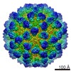 5410C 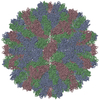 3j1pC  4ejrC  2gh8S C: citing same article ( S: Starting model for refinement |
|---|---|
| Similar structure data |
-Links
 PDBj
PDBj

- Assembly
| Deposited unit | 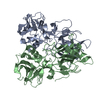
| ||||||||
|---|---|---|---|---|---|---|---|---|---|
| 1 |
| ||||||||
| Unit cell |
|
-Components
| #1: Protein | Mass: 37067.852 Da / Num. of mol.: 2 / Fragment: P (protruding) domain (UNP residues 228-579) Source method: isolated from a genetically manipulated source Source: (gene. exp.) Rabbit hemorrhagic disease virus / Gene: VP60 / Plasmid: pFastEL-3G / Production host:   Spodoptera frugiperda (fall armyworm) / Strain (production host): Sf21 / References: UniProt: Q3HNQ1, UniProt: Q3HNQ2*PLUS Spodoptera frugiperda (fall armyworm) / Strain (production host): Sf21 / References: UniProt: Q3HNQ1, UniProt: Q3HNQ2*PLUS#2: Water | ChemComp-HOH / |  Mass: 18.015 Da / Num. of mol.: 192 / Source method: isolated from a natural source / Formula: H2O Mass: 18.015 Da / Num. of mol.: 192 / Source method: isolated from a natural source / Formula: H2OSequence details | THE AUTHORS STATE THAT G263R AND G539E ARE NATURAL SEQUENCE VARIATIONS | |
|---|
-Experimental details
-Experiment
| Experiment | Method: X-RAY DIFFRACTION / Number of used crystals: 1 |
|---|
- Sample preparation
| Crystal | Density Matthews: 1.77 Å3/Da / Density % sol: 30.53 % |
|---|---|
| Crystal grow | Temperature: 289 K / Method: vapor diffusion / pH: 5.5 Details: 0.1 M sodium acetate, 1.1 M succinic acid, 1.0% PEG2000 MME, pH 5.5, VAPOR DIFFUSION, temperature 289K |
-Data collection
| Diffraction | Mean temperature: 100 K |
|---|---|
| Diffraction source | Source: SYNCHROTRON / Site: SSRF / Beamline: BL17U / Wavelength: 0.99985 Å |
| Detector | Type: RAYONIX MX-225 / Detector: CCD / Date: Oct 21, 2010 |
| Radiation | Monochromator: double crystal Si(111) / Protocol: SINGLE WAVELENGTH / Monochromatic (M) / Laue (L): M / Scattering type: x-ray |
| Radiation wavelength | Wavelength: 0.99985 Å / Relative weight: 1 |
| Reflection | Resolution: 2→50 Å / Num. all: 36207 / Num. obs: 35882 / % possible obs: 99.1 % / Observed criterion σ(F): 0 / Observed criterion σ(I): -2 / Redundancy: 3.7 % / Rmerge(I) obs: 0.089 / Net I/σ(I): 29.8 |
| Reflection shell | Resolution: 2→2.03 Å / Redundancy: 3.9 % / Rmerge(I) obs: 0.231 / Mean I/σ(I) obs: 8.3 / Num. unique all: 1727 / % possible all: 99.9 |
- Processing
| Software |
| ||||||||||||||||||||||||||||||||||||||||||||||||||||||||||||||||||||||||||||||||||||||||||||||||||||||||||||||||||||||||||||||||||||||||||||||||||||||||||||||||||||||||||
|---|---|---|---|---|---|---|---|---|---|---|---|---|---|---|---|---|---|---|---|---|---|---|---|---|---|---|---|---|---|---|---|---|---|---|---|---|---|---|---|---|---|---|---|---|---|---|---|---|---|---|---|---|---|---|---|---|---|---|---|---|---|---|---|---|---|---|---|---|---|---|---|---|---|---|---|---|---|---|---|---|---|---|---|---|---|---|---|---|---|---|---|---|---|---|---|---|---|---|---|---|---|---|---|---|---|---|---|---|---|---|---|---|---|---|---|---|---|---|---|---|---|---|---|---|---|---|---|---|---|---|---|---|---|---|---|---|---|---|---|---|---|---|---|---|---|---|---|---|---|---|---|---|---|---|---|---|---|---|---|---|---|---|---|---|---|---|---|---|---|---|---|
| Refinement | Method to determine structure: MOLECULAR REPLACEMENT Starting model: PDB ENTRY 2GH8 Resolution: 2→36.95 Å / Cor.coef. Fo:Fc: 0.95 / Cor.coef. Fo:Fc free: 0.928 / Cross valid method: THROUGHOUT / σ(F): 0 / ESU R: 0.248 / ESU R Free: 0.183 / Stereochemistry target values: MAXIMUM LIKELIHOOD Details: HYDROGENS HAVE BEEN ADDED IN THE RIDING POSITIONS U VALUES : REFINED INDIVIDUALLY
| ||||||||||||||||||||||||||||||||||||||||||||||||||||||||||||||||||||||||||||||||||||||||||||||||||||||||||||||||||||||||||||||||||||||||||||||||||||||||||||||||||||||||||
| Solvent computation | Ion probe radii: 0.8 Å / Shrinkage radii: 0.8 Å / VDW probe radii: 1.4 Å / Solvent model: MASK | ||||||||||||||||||||||||||||||||||||||||||||||||||||||||||||||||||||||||||||||||||||||||||||||||||||||||||||||||||||||||||||||||||||||||||||||||||||||||||||||||||||||||||
| Displacement parameters | Biso mean: 22.745 Å2
| ||||||||||||||||||||||||||||||||||||||||||||||||||||||||||||||||||||||||||||||||||||||||||||||||||||||||||||||||||||||||||||||||||||||||||||||||||||||||||||||||||||||||||
| Refinement step | Cycle: LAST / Resolution: 2→36.95 Å
| ||||||||||||||||||||||||||||||||||||||||||||||||||||||||||||||||||||||||||||||||||||||||||||||||||||||||||||||||||||||||||||||||||||||||||||||||||||||||||||||||||||||||||
| Refine LS restraints |
| ||||||||||||||||||||||||||||||||||||||||||||||||||||||||||||||||||||||||||||||||||||||||||||||||||||||||||||||||||||||||||||||||||||||||||||||||||||||||||||||||||||||||||
| LS refinement shell | Resolution: 2→2.057 Å / Total num. of bins used: 20
|
