 Movie
Movie Controller

[English] 日本語
 Yorodumi
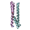
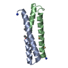
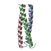
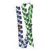
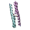
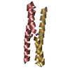
- PDB-7bey: Het-N2-SO3- - De novo designed three-helix heterodimer with Cyste... -
+ Open data

- Basic information
| Entry | Database: PDB / ID: 7bey | ||||||||||||
|---|---|---|---|---|---|---|---|---|---|---|---|---|---|
| Title | Het-N2-SO3- - De novo designed three-helix heterodimer with Cysteine S-sulfate at the N2 position of the alpha-helix | ||||||||||||
 Components Components |
| ||||||||||||
 Keywords Keywords | DE NOVO PROTEIN / Acyl Transfer Activity / Domain Swapped Dimer / Oxyanion-Binding Site | ||||||||||||
| Biological species | synthetic construct (others) | ||||||||||||
| Method |  X-RAY DIFFRACTION / SYNCHROTRON / MOLECULAR REPLACEMENT / molecular replacement / Resolution: 1.5 Å X-RAY DIFFRACTION / SYNCHROTRON / MOLECULAR REPLACEMENT / molecular replacement / Resolution: 1.5 Å | ||||||||||||
 Authors Authors | McEwen, A.G. / Poussin-Courmontagne, P. / Naudin, E.A. / DeGrado, W.F. / Torbeev, V. | ||||||||||||
| Funding support | European Union,  France, 3items France, 3items
| ||||||||||||
 Citation Citation | Journal: J.Am.Chem.Soc. / Year: 2021 Title: Acyl Transfer Catalytic Activity in De Novo Designed Protein with N-Terminus of alpha-Helix As Oxyanion-Binding Site. Authors: Naudin, E.A. / McEwen, A.G. / Tan, S.K. / Poussin-Courmontagne, P. / Schmitt, J.L. / Birck, C. / DeGrado, W.F. / Torbeev, V. | ||||||||||||
| History |
|
- Structure visualization
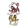
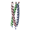
| Structure viewer | Molecule:  MolmilJmol/JSmol MolmilJmol/JSmol |
|---|
- Downloads & links
-Download
| PDBx/mmCIF format | 7bey.cif.gz | 135.3 KB | Display | PDBx/mmCIF format |
|---|---|---|---|---|
| PDB format | pdb7bey.ent.gz | 111.4 KB | Display | PDB format |
| PDBx/mmJSON format | 7bey.json.gz | Tree view | PDBx/mmJSON format | |
| Others |  Other downloads Other downloads |
-Validation report
| Arichive directory | https://data.pdbj.org/pub/pdb/validation_reports/be/7beyftp://data.pdbj.org/pub/pdb/validation_reports/be/7bey | HTTPS FTP |
|---|
-Related structure data
| Related structure data |  6z0lSC 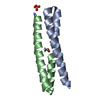 6z0mC S: Starting model for refinement C: citing same article ( |
|---|---|
| Similar structure data |

-Links
 PDBj
PDBj
- Assembly
| Deposited unit | 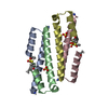
| ||||||||
|---|---|---|---|---|---|---|---|---|---|
| 1 | 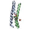
| ||||||||
| 2 | 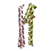
| ||||||||
| Unit cell |
|
-Components
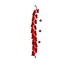
| #1: Protein/peptide | Mass: 5462.353 Da / Num. of mol.: 2 / Source method: obtained synthetically / Source: (synth.) synthetic construct (others) #2: Protein/peptide | Mass: 5405.109 Da / Num. of mol.: 2 / Source method: obtained synthetically / Source: (synth.) synthetic construct (others) #3: Chemical |   Mass: 96.063 Da / Num. of mol.: 2 / Source method: obtained synthetically / Formula: SO4 Mass: 96.063 Da / Num. of mol.: 2 / Source method: obtained synthetically / Formula: SO4#4: Water | ChemComp-HOH / |  Mass: 18.015 Da / Num. of mol.: 238 / Source method: isolated from a natural source / Formula: H2O Mass: 18.015 Da / Num. of mol.: 238 / Source method: isolated from a natural source / Formula: H2OHas ligand of interest | Y | |
|---|
-Experimental details
-Experiment
| Experiment | Method: X-RAY DIFFRACTION / Number of used crystals: 1 |
|---|
- Sample preparation
| Crystal | Density Matthews: 1.95 Å3/Da / Density % sol: 37.08 % |
|---|---|
| Crystal grow | Temperature: 293 K / Method: vapor diffusion, sitting drop / pH: 4.2 Details: 10% propan-2-ol, 0.2 M lithium sulfate, 0.1 M sodium phosphate citrate pH 4.2 |
-Data collection
| Diffraction | Mean temperature: 100 K / Serial crystal experiment: N | ||||||||||||||||||||||||||||||||||||||||||||||||||||||||||||||||||||||||||||||||||||||||||||||||||||
|---|---|---|---|---|---|---|---|---|---|---|---|---|---|---|---|---|---|---|---|---|---|---|---|---|---|---|---|---|---|---|---|---|---|---|---|---|---|---|---|---|---|---|---|---|---|---|---|---|---|---|---|---|---|---|---|---|---|---|---|---|---|---|---|---|---|---|---|---|---|---|---|---|---|---|---|---|---|---|---|---|---|---|---|---|---|---|---|---|---|---|---|---|---|---|---|---|---|---|---|---|---|
| Diffraction source | Source: SYNCHROTRON / Site: SOLEIL / Beamline: PROXIMA 1 / Wavelength: 0.979 Å | ||||||||||||||||||||||||||||||||||||||||||||||||||||||||||||||||||||||||||||||||||||||||||||||||||||
| Detector | Type: DECTRIS EIGER X 16M / Detector: PIXEL / Date: Oct 9, 2020 | ||||||||||||||||||||||||||||||||||||||||||||||||||||||||||||||||||||||||||||||||||||||||||||||||||||
| Radiation | Monochromator: Si(111) / Protocol: SINGLE WAVELENGTH / Monochromatic (M) / Laue (L): M / Scattering type: x-ray | ||||||||||||||||||||||||||||||||||||||||||||||||||||||||||||||||||||||||||||||||||||||||||||||||||||
| Radiation wavelength | Wavelength: 0.979 Å / Relative weight: 1 | ||||||||||||||||||||||||||||||||||||||||||||||||||||||||||||||||||||||||||||||||||||||||||||||||||||
| Reflection | Resolution: 1.5→34.34 Å / Num. obs: 26503 / % possible obs: 98.5 % / Redundancy: 6.949 % / Biso Wilson estimate: 29.255 Å2 / CC1/2: 0.998 / Rmerge(I) obs: 0.195 / Rrim(I) all: 0.211 / Χ2: 0.736 / Net I/σ(I): 5.34 / Num. measured all: 184181 | ||||||||||||||||||||||||||||||||||||||||||||||||||||||||||||||||||||||||||||||||||||||||||||||||||||
| Reflection shell | Diffraction-ID: 1
|
-Phasing
| Phasing | Method: molecular replacement | |||||||||
|---|---|---|---|---|---|---|---|---|---|---|
| Phasing MR |
|
- Processing
| Software |
| ||||||||||||||||||||||||||||||||||||||||||
|---|---|---|---|---|---|---|---|---|---|---|---|---|---|---|---|---|---|---|---|---|---|---|---|---|---|---|---|---|---|---|---|---|---|---|---|---|---|---|---|---|---|---|---|
| Refinement | Method to determine structure: MOLECULAR REPLACEMENT Starting model: 6Z0L Resolution: 1.5→34.34 Å / SU ML: 0.23 / Cross valid method: THROUGHOUT / σ(F): 1.35 / Phase error: 38.64 / Stereochemistry target values: ML
| ||||||||||||||||||||||||||||||||||||||||||
| Solvent computation | Shrinkage radii: 0.9 Å / VDW probe radii: 1.11 Å / Solvent model: FLAT BULK SOLVENT MODEL | ||||||||||||||||||||||||||||||||||||||||||
| Displacement parameters | Biso max: 91.94 Å2 / Biso mean: 18.7681 Å2 / Biso min: 6.56 Å2 | ||||||||||||||||||||||||||||||||||||||||||
| Refinement step | Cycle: final / Resolution: 1.5→34.34 Å
| ||||||||||||||||||||||||||||||||||||||||||
| LS refinement shell | Refine-ID: X-RAY DIFFRACTION / Rfactor Rfree error: 0 / Total num. of bins used: 5
|
