 Movie
Movie Controller

[English] 日本語
 Yorodumi
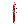
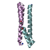
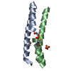
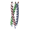
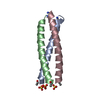
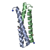
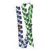
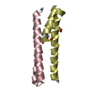
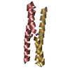
- PDB-1ce0: TRIMERIZATION SPECIFICITY IN HIV-1 GP41: ANALYSIS WITH A GCN4 LEU... -
+ Open data

- Basic information
| Entry | Database: PDB / ID: 1ce0 | ||||||
|---|---|---|---|---|---|---|---|
| Title | TRIMERIZATION SPECIFICITY IN HIV-1 GP41: ANALYSIS WITH A GCN4 LEUCINE ZIPPER MODEL | ||||||
 Components Components | PROTEIN (LEUCINE ZIPPER MODEL H38-P1) | ||||||
 Keywords Keywords | HIV-1 ENVELOPE PROTEIN / GP41 / PROTEIN OLIGOMERIZATION / COILED COIL / LEUCINE ZIPPER | ||||||
| Function / homology | :  Function and homology information Function and homology information | ||||||
| Biological species |   Human immunodeficiency virus 1 Human immunodeficiency virus 1 | ||||||
| Method |  X-RAY DIFFRACTION / SYNCHROTRON / MOLECULAR REPLACEMENT / Resolution: 2.4 Å X-RAY DIFFRACTION / SYNCHROTRON / MOLECULAR REPLACEMENT / Resolution: 2.4 Å | ||||||
 Authors Authors | Shu, W. / Ji, H. / Lu, M. | ||||||
 Citation Citation | Journal: Biochemistry / Year: 1999 Title: Trimerization specificity in HIV-1 gp41: analysis with a GCN4 leucine zipper model. Authors: Shu, W. / Ji, H. / Lu, M. #1: Journal: Science / Year: 1991Title: X-ray structure of the GCN4 leucine zipper, a two-stranded, parallel coiled coil. Authors: O'Shea, E.K. / Klemm, J.D. / Kim, P.S. / Alber, T. | ||||||
| History |
|
- Structure visualization
| Structure viewer | Molecule: MolmilJmol/JSmol |
|---|
- Downloads & links
-Download
| PDBx/mmCIF format | 1ce0.cif.gz | 33.3 KB | Display | PDBx/mmCIF format |
|---|---|---|---|---|
| PDB format | pdb1ce0.ent.gz | 24.3 KB | Display | PDB format |
| PDBx/mmJSON format | 1ce0.json.gz | Tree view | PDBx/mmJSON format | |
| Others |  Other downloads Other downloads |
-Validation report
| Arichive directory | https://data.pdbj.org/pub/pdb/validation_reports/ce/1ce0ftp://data.pdbj.org/pub/pdb/validation_reports/ce/1ce0 | HTTPS FTP |
|---|
-Related structure data
| Related structure data | 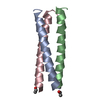 1gcmS S: Starting model for refinement |
|---|---|
| Similar structure data |

-Links
 PDBj
PDBj- Assembly
| Deposited unit | 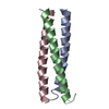
| ||||||||||
|---|---|---|---|---|---|---|---|---|---|---|---|
| 1 |
| ||||||||||
| Unit cell |
|
-Components
| #1: Protein/peptide | Mass: 4529.289 Da / Num. of mol.: 3 Source method: isolated from a genetically manipulated source Source: (gene. exp.) Human immunodeficiency virus 1 / Genus: Lentivirus / Description: SYNTHETIC GENE / Plasmid: PH38-P1 / Production host:  #2: Water | ChemComp-HOH / |  Mass: 18.015 Da / Num. of mol.: 78 / Source method: isolated from a natural source / Formula: H2O Mass: 18.015 Da / Num. of mol.: 78 / Source method: isolated from a natural source / Formula: H2O |
|---|
-Experimental details
-Experiment
| Experiment | Method: X-RAY DIFFRACTION / Number of used crystals: 1 |
|---|
- Sample preparation
| Crystal | Density Matthews: 3.27 Å3/Da / Density % sol: 69.7 % | ||||||||||||||||||||||||||||||||||||
|---|---|---|---|---|---|---|---|---|---|---|---|---|---|---|---|---|---|---|---|---|---|---|---|---|---|---|---|---|---|---|---|---|---|---|---|---|---|
| Crystal grow | pH: 7.5 / Details: pH 7.5 | ||||||||||||||||||||||||||||||||||||
| Crystal grow | *PLUS Method: vapor diffusion, hanging drop | ||||||||||||||||||||||||||||||||||||
| Components of the solutions | *PLUS
|
-Data collection
| Diffraction | Mean temperature: 100 K |
|---|---|
| Diffraction source | Source: SYNCHROTRON / Site: NSLS  / Beamline: X12B / Wavelength: 0.978 / Beamline: X12B / Wavelength: 0.978 |
| Detector | Type: MARRESEARCH / Detector: IMAGE PLATE / Date: Oct 15, 1998 / Details: REFOCUSSING MIRROR, RH WORKING SURFACE |
| Radiation | Monochromator: DOUBLE-CRYSTAL, FIXED-EXIT SI-III MONOCHROMATOR Protocol: SINGLE WAVELENGTH / Monochromatic (M) / Laue (L): M / Scattering type: x-ray |
| Radiation wavelength | Wavelength: 0.978 Å / Relative weight: 1 |
| Reflection | Resolution: 2.4→30 Å / Num. obs: 6765 / % possible obs: 98.8 % / Observed criterion σ(I): -3 / Redundancy: 3.3 % / Rmerge(I) obs: 0.039 / Rsym value: 0.039 / Net I/σ(I): 20.4 |
| Reflection shell | Resolution: 2.4→2.49 Å / Redundancy: 3.3 % / Rmerge(I) obs: 0.097 / Mean I/σ(I) obs: 10.5 / Rsym value: 0.097 / % possible all: 99.8 |
| Reflection | *PLUS Num. measured all: 22836 |
- Processing
| Software |
| ||||||||||||||||||||||||||||||||||||||||||||||||||||||||||||
|---|---|---|---|---|---|---|---|---|---|---|---|---|---|---|---|---|---|---|---|---|---|---|---|---|---|---|---|---|---|---|---|---|---|---|---|---|---|---|---|---|---|---|---|---|---|---|---|---|---|---|---|---|---|---|---|---|---|---|---|---|---|
| Refinement | Method to determine structure: MOLECULAR REPLACEMENT Starting model: PDB ENTRY 1GCM Resolution: 2.4→8 Å / Cross valid method: THROUGHOUT / σ(F): 2
| ||||||||||||||||||||||||||||||||||||||||||||||||||||||||||||
| Displacement parameters | Biso mean: 37.5 Å2 | ||||||||||||||||||||||||||||||||||||||||||||||||||||||||||||
| Refinement step | Cycle: LAST / Resolution: 2.4→8 Å
| ||||||||||||||||||||||||||||||||||||||||||||||||||||||||||||
| Refine LS restraints |
| ||||||||||||||||||||||||||||||||||||||||||||||||||||||||||||
| Software | *PLUS Name: X-PLOR / Version: 3.851 / Classification: refinement | ||||||||||||||||||||||||||||||||||||||||||||||||||||||||||||
| Refinement | *PLUS Highest resolution: 2.4 Å / Lowest resolution: 8 Å / σ(F): 2 / % reflection Rfree: 7 % / Rfactor obs: 0.221 | ||||||||||||||||||||||||||||||||||||||||||||||||||||||||||||
| Solvent computation | *PLUS | ||||||||||||||||||||||||||||||||||||||||||||||||||||||||||||
| Displacement parameters | *PLUS Biso mean: 37.5 Å2 | ||||||||||||||||||||||||||||||||||||||||||||||||||||||||||||
| Refine LS restraints | *PLUS
|
