 Movie
Movie Controller


+ Open data

- Basic information
| Entry | Database: PDB / ID: 7bcc | ||||||
|---|---|---|---|---|---|---|---|
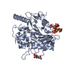
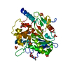
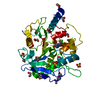
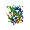
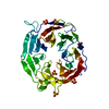
| Title | Notum Fragment 705 | ||||||
 Components Components | Palmitoleoyl-protein carboxylesterase NOTUM | ||||||
 Keywords Keywords | HYDROLASE / Notum Inhibitor | ||||||
| Function / homology |  Function and homology information Function and homology information[Wnt protein] O-palmitoleoyl-L-serine hydrolase / protein-containing complex destabilizing activity / protein depalmitoleylation / palmitoleyl hydrolase activity / Release of Hh-Np from the secreting cell / C-type glycerophospholipase activity / regulation of bone mineralization / negative regulation of Wnt signaling pathway / Post-translational protein phosphorylation / negative regulation of canonical Wnt signaling pathway ...[Wnt protein] O-palmitoleoyl-L-serine hydrolase / protein-containing complex destabilizing activity / protein depalmitoleylation / palmitoleyl hydrolase activity / Release of Hh-Np from the secreting cell / C-type glycerophospholipase activity / regulation of bone mineralization / negative regulation of Wnt signaling pathway / Post-translational protein phosphorylation / negative regulation of canonical Wnt signaling pathway / bone development / Regulation of Insulin-like Growth Factor (IGF) transport and uptake by Insulin-like Growth Factor Binding Proteins (IGFBPs) / Wnt signaling pathway / endoplasmic reticulum lumen / extracellular region Similarity search - Function | ||||||
| Biological species |  Homo sapiens (human) Homo sapiens (human) | ||||||
| Method |  X-RAY DIFFRACTION / SYNCHROTRON / MOLECULAR REPLACEMENT / Resolution: 1.58 Å X-RAY DIFFRACTION / SYNCHROTRON / MOLECULAR REPLACEMENT / Resolution: 1.58 Å | ||||||
 Authors Authors | Zhao, Y. / Jones, E.Y. | ||||||
| Funding support |  United Kingdom, 1items United Kingdom, 1items
| ||||||
 Citation Citation | Journal: Acs Chem Neurosci / Year: 2022 Title: Structural Analysis and Development of Notum Fragment Screening Hits. Authors: Zhao, Y. / Mahy, W. / Willis, N.J. / Woodward, H.L. / Steadman, D. / Bayle, E.D. / Atkinson, B.N. / Sipthorp, J. / Vecchia, L. / Ruza, R.R. / Harlos, K. / Jeganathan, F. / Constantinou, S. / ...Authors: Zhao, Y. / Mahy, W. / Willis, N.J. / Woodward, H.L. / Steadman, D. / Bayle, E.D. / Atkinson, B.N. / Sipthorp, J. / Vecchia, L. / Ruza, R.R. / Harlos, K. / Jeganathan, F. / Constantinou, S. / Costa, A. / Kjaer, S. / Bictash, M. / Salinas, P.C. / Whiting, P. / Vincent, J.P. / Fish, P.V. / Jones, E.Y. | ||||||
| History |
|
- Structure visualization
| Structure viewer | Molecule: MolmilJmol/JSmol |
|---|
- Downloads & links
-Download
| PDBx/mmCIF format | 7bcc.cif.gz | 161.6 KB | Display | PDBx/mmCIF format |
|---|---|---|---|---|
| PDB format | pdb7bcc.ent.gz | 123.9 KB | Display | PDB format |
| PDBx/mmJSON format | 7bcc.json.gz | Tree view | PDBx/mmJSON format | |
| Others |  Other downloads Other downloads |
-Validation report
| Arichive directory | https://data.pdbj.org/pub/pdb/validation_reports/bc/7bccftp://data.pdbj.org/pub/pdb/validation_reports/bc/7bcc | HTTPS FTP |
|---|
-Related structure data
| Related structure data | 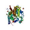 7b4xC 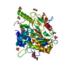 7b84C 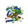 7b86C 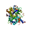 7b87C  7b89C 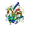 7b8cC 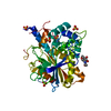 7b8dC 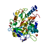 7b8fC 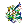 7b8gC 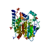 7b8jC 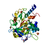 7b8kC  7b8lC 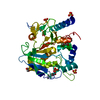 7b8mC  7b8nC  7b8oC 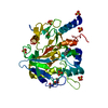 7b8uC  7b8xC 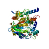 7b8yC 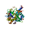 7b8zC 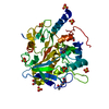 7b98C 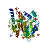 7b99C 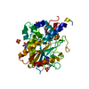 7b9dC 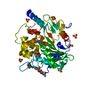 7b9iC  7b9nC 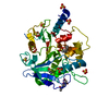 7b9uC 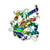 7ba1C 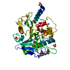 7bacC 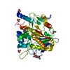 7bapC 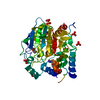 7bc8C 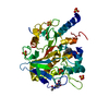 7bc9C 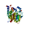 7bcdC  7bceC 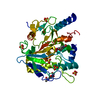 7bcfC 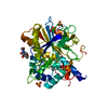 7bchC 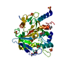 7bciC  7bckC 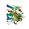 7bclC 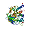 7bd2C 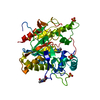 7bd3C 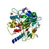 7bd4C  7bd5C 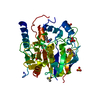 7bd6C 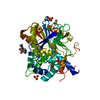 7bd8C 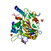 7bd9C 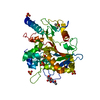 7bdaC  7bdbC 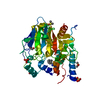 7bdcC 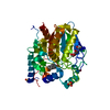 7bddC  7bdfC  7bdgC 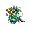 7bdhC  4uzqS C: citing same article ( S: Starting model for refinement |
|---|---|
| Similar structure data |


-Links
 PDBj
PDBj- Assembly
| Deposited unit | 
| ||||||||
|---|---|---|---|---|---|---|---|---|---|
| 1 |
| ||||||||
| Unit cell |
|
-Components
| #1: Protein | Mass: 43567.148 Da / Num. of mol.: 1 Source method: isolated from a genetically manipulated source Source: (gene. exp.) Homo sapiens (human) / Gene: NOTUM, OK/SW-CL.30 / Production host: Homo sapiens (human)References: UniProt: Q6P988, [Wnt protein] O-palmitoleoyl-L-serine hydrolase | ||||||||||
|---|---|---|---|---|---|---|---|---|---|---|---|
| #2: Chemical | ChemComp-SO4 /   Mass: 96.063 Da / Num. of mol.: 5 / Source method: obtained synthetically / Formula: SO4 Mass: 96.063 Da / Num. of mol.: 5 / Source method: obtained synthetically / Formula: SO4#3: Sugar | ChemComp-NAG / |   Type: D-saccharide, beta linking / Mass: 221.208 Da / Num. of mol.: 1 / Source method: obtained synthetically / Formula: C8H15NO6 Type: D-saccharide, beta linking / Mass: 221.208 Da / Num. of mol.: 1 / Source method: obtained synthetically / Formula: C8H15NO6#4: Chemical | ChemComp-TB5 / |   Mass: 228.290 Da / Num. of mol.: 1 / Source method: obtained synthetically / Formula: C14H16N2O / Feature type: SUBJECT OF INVESTIGATION Mass: 228.290 Da / Num. of mol.: 1 / Source method: obtained synthetically / Formula: C14H16N2O / Feature type: SUBJECT OF INVESTIGATION#5: Water | ChemComp-HOH / |  Mass: 18.015 Da / Num. of mol.: 92 / Source method: isolated from a natural source / Formula: H2O Mass: 18.015 Da / Num. of mol.: 92 / Source method: isolated from a natural source / Formula: H2OHas ligand of interest | Y | Has protein modification | Y | |
-Experimental details
-Experiment
| Experiment | Method: X-RAY DIFFRACTION / Number of used crystals: 1 |
|---|
- Sample preparation
| Crystal | Density Matthews: 1.98 Å3/Da / Density % sol: 38.02 % |
|---|---|
| Crystal grow | Temperature: 296 K / Method: vapor diffusion, sitting drop Details: 1.5 M Ammonium Sulphate 0.1 M Sodium Citrate, pH4.2 |
-Data collection
| Diffraction | Mean temperature: 100 K / Serial crystal experiment: N |
|---|---|
| Diffraction source | Source: SYNCHROTRON / Site: Diamond / Beamline: I04-1 / Wavelength: 0.9282 Å |
| Detector | Type: DECTRIS PILATUS3 S 6M / Detector: PIXEL / Date: Feb 2, 2017 |
| Radiation | Protocol: SINGLE WAVELENGTH / Monochromatic (M) / Laue (L): M / Scattering type: x-ray |
| Radiation wavelength | Wavelength: 0.9282 Å / Relative weight: 1 |
| Reflection | Resolution: 1.58→78.87 Å / Num. obs: 48153 / % possible obs: 100 % / Redundancy: 6.3 % / CC1/2: 1 / Net I/σ(I): 9.2 |
| Reflection shell | Resolution: 1.58→1.61 Å / Num. unique obs: 2327 / CC1/2: 0.7 |
- Processing
| Software |
| ||||||||||||||||||||||||||||||||||||||||||||||||||||||||||||||||||||||||||||||||||||||||||||||||||||||||||||||||||||||||||||||
|---|---|---|---|---|---|---|---|---|---|---|---|---|---|---|---|---|---|---|---|---|---|---|---|---|---|---|---|---|---|---|---|---|---|---|---|---|---|---|---|---|---|---|---|---|---|---|---|---|---|---|---|---|---|---|---|---|---|---|---|---|---|---|---|---|---|---|---|---|---|---|---|---|---|---|---|---|---|---|---|---|---|---|---|---|---|---|---|---|---|---|---|---|---|---|---|---|---|---|---|---|---|---|---|---|---|---|---|---|---|---|---|---|---|---|---|---|---|---|---|---|---|---|---|---|---|---|---|
| Refinement | Method to determine structure: MOLECULAR REPLACEMENT Starting model: 4UZQ Resolution: 1.58→53.38 Å / SU ML: 0.24 / Cross valid method: THROUGHOUT / σ(F): 1.33 / Phase error: 28.13 / Stereochemistry target values: ML
| ||||||||||||||||||||||||||||||||||||||||||||||||||||||||||||||||||||||||||||||||||||||||||||||||||||||||||||||||||||||||||||||
| Solvent computation | Shrinkage radii: 0.9 Å / VDW probe radii: 1.11 Å / Solvent model: FLAT BULK SOLVENT MODEL | ||||||||||||||||||||||||||||||||||||||||||||||||||||||||||||||||||||||||||||||||||||||||||||||||||||||||||||||||||||||||||||||
| Displacement parameters | Biso max: 59.45 Å2 / Biso mean: 25.0381 Å2 / Biso min: 13.39 Å2 | ||||||||||||||||||||||||||||||||||||||||||||||||||||||||||||||||||||||||||||||||||||||||||||||||||||||||||||||||||||||||||||||
| Refinement step | Cycle: final / Resolution: 1.58→53.38 Å
| ||||||||||||||||||||||||||||||||||||||||||||||||||||||||||||||||||||||||||||||||||||||||||||||||||||||||||||||||||||||||||||||
| LS refinement shell | Refine-ID: X-RAY DIFFRACTION / Rfactor Rfree error: 0 / Total num. of bins used: 17
| ||||||||||||||||||||||||||||||||||||||||||||||||||||||||||||||||||||||||||||||||||||||||||||||||||||||||||||||||||||||||||||||
| Refinement TLS params. | Method: refined / Origin x: 4.46 Å / Origin y: -1.241 Å / Origin z: -1.948 Å
| ||||||||||||||||||||||||||||||||||||||||||||||||||||||||||||||||||||||||||||||||||||||||||||||||||||||||||||||||||||||||||||||
| Refinement TLS group |
|