 Movie
Movie Controller


+ Open data

- Basic information
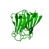
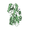
| Entry | Database: PDB / ID: 7b63 | ||||||
|---|---|---|---|---|---|---|---|
| Title | Structure of NUDT15 in complex with TH7755 | ||||||
 Components Components | Probable 8-oxo-dGTP diphosphatase NUDT15 | ||||||
 Keywords Keywords | HYDROLASE / Inhibitor / complex / Nucleoside Triphosphate Pyrophosphohydrolase | ||||||
| Function / homology |  Function and homology information Function and homology informationnucleoside phosphate catabolic process / prenyl-diphosphate phosphatase / isoprenoid diphosphate phosphatase activity / geranyl diphosphate phosphohydrolase / purine nucleotide catabolic process / nucleotide diphosphatase / nucleobase-containing small molecule metabolic process / nucleoside triphosphate diphosphatase activity / 8-oxo-dGDP phosphatase activity / dGTP catabolic process ...nucleoside phosphate catabolic process / prenyl-diphosphate phosphatase / isoprenoid diphosphate phosphatase activity / geranyl diphosphate phosphohydrolase / purine nucleotide catabolic process / nucleotide diphosphatase / nucleobase-containing small molecule metabolic process / nucleoside triphosphate diphosphatase activity / 8-oxo-dGDP phosphatase activity / dGTP catabolic process / 8-oxo-7,8-dihydrodeoxyguanosine triphosphate pyrophosphatase activity / 8-oxo-7,8-dihydroguanosine triphosphate pyrophosphatase activity / DNA protection / Phosphate bond hydrolysis by NUDT proteins / Azathioprine ADME / xenobiotic catabolic process / regulation of proteasomal protein catabolic process / response to reactive oxygen species / mitotic cell cycle / metal ion binding / cytosol Similarity search - Function | ||||||
| Biological species |  Homo sapiens (human) Homo sapiens (human) | ||||||
| Method |  X-RAY DIFFRACTION / SYNCHROTRON / MOLECULAR REPLACEMENT / Resolution: 1.6 Å X-RAY DIFFRACTION / SYNCHROTRON / MOLECULAR REPLACEMENT / Resolution: 1.6 Å | ||||||
 Authors Authors | Rehling, D. / Stenmark, P. | ||||||
| Funding support |  Sweden, 1items Sweden, 1items
| ||||||
 Citation Citation | Journal: J.Biol.Chem. / Year: 2021 Title: Crystal structures of NUDT15 variants enabled by a potent inhibitor reveal the structural basis for thiopurine sensitivity. Authors: Rehling, D. / Zhang, S.M. / Jemth, A.S. / Koolmeister, T. / Throup, A. / Wallner, O. / Scaletti, E. / Moriyama, T. / Nishii, R. / Davies, J. / Desroses, M. / Rudd, S.G. / Scobie, M. / Homan, ...Authors: Rehling, D. / Zhang, S.M. / Jemth, A.S. / Koolmeister, T. / Throup, A. / Wallner, O. / Scaletti, E. / Moriyama, T. / Nishii, R. / Davies, J. / Desroses, M. / Rudd, S.G. / Scobie, M. / Homan, E. / Berglund, U.W. / Yang, J.J. / Helleday, T. / Stenmark, P. | ||||||
| History |
|
- Structure visualization
| Structure viewer | Molecule: MolmilJmol/JSmol |
|---|
- Downloads & links
-Download
| PDBx/mmCIF format | 7b63.cif.gz | 89.8 KB | Display | PDBx/mmCIF format |
|---|---|---|---|---|
| PDB format | pdb7b63.ent.gz | 63.8 KB | Display | PDB format |
| PDBx/mmJSON format | 7b63.json.gz | Tree view | PDBx/mmJSON format | |
| Others |  Other downloads Other downloads |
-Validation report
| Arichive directory | https://data.pdbj.org/pub/pdb/validation_reports/b6/7b63ftp://data.pdbj.org/pub/pdb/validation_reports/b6/7b63 | HTTPS FTP |
|---|
-Related structure data
| Related structure data | 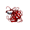 7b64C 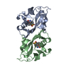 7b65C 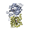 7b66C  7b67C 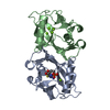 5lpgS S: Starting model for refinement C: citing same article ( |
|---|---|
| Similar structure data |


-Links
 PDBj
PDBj

- Assembly
| Deposited unit | 
| ||||||||
|---|---|---|---|---|---|---|---|---|---|
| 1 |
| ||||||||
| Unit cell |
|
-Components
| #1: Protein | Mass: 18635.002 Da / Num. of mol.: 2 Source method: isolated from a genetically manipulated source Source: (gene. exp.) Homo sapiens (human) / Gene: NUDT15, MTH2 / Production host:  #2: Chemical |   Mass: 454.499 Da / Num. of mol.: 2 / Source method: obtained synthetically / Formula: C22H22N4O5S / Feature type: SUBJECT OF INVESTIGATION Mass: 454.499 Da / Num. of mol.: 2 / Source method: obtained synthetically / Formula: C22H22N4O5S / Feature type: SUBJECT OF INVESTIGATION#3: Chemical | ChemComp-MG / |   Mass: 24.305 Da / Num. of mol.: 1 / Source method: obtained synthetically / Formula: Mg Mass: 24.305 Da / Num. of mol.: 1 / Source method: obtained synthetically / Formula: Mg#4: Water | ChemComp-HOH / |  Mass: 18.015 Da / Num. of mol.: 348 / Source method: isolated from a natural source / Formula: H2O Mass: 18.015 Da / Num. of mol.: 348 / Source method: isolated from a natural source / Formula: H2OHas ligand of interest | Y | |
|---|
-Experimental details
-Experiment
| Experiment | Method: X-RAY DIFFRACTION / Number of used crystals: 1 |
|---|
- Sample preparation
| Crystal | Density Matthews: 2.35 Å3/Da / Density % sol: 47.57 % |
|---|---|
| Crystal grow | Temperature: 294 K / Method: vapor diffusion, sitting drop Details: 0.1 M Tris pH 8.0, 0.2 M sodium acetate, 38% PEG 4000 |
-Data collection
| Diffraction | Mean temperature: 100 K / Serial crystal experiment: N |
|---|---|
| Diffraction source | Source: SYNCHROTRON / Site: MAX IV / Beamline: BioMAX / Wavelength: 0.9184 Å |
| Detector | Type: DECTRIS EIGER X 16M / Detector: PIXEL / Date: Feb 20, 2019 |
| Radiation | Protocol: SINGLE WAVELENGTH / Monochromatic (M) / Laue (L): M / Scattering type: x-ray |
| Radiation wavelength | Wavelength: 0.9184 Å / Relative weight: 1 |
| Reflection | Resolution: 1.6→60.24 Å / Num. obs: 45919 / % possible obs: 100 % / Redundancy: 6.8 % / CC1/2: 0.999 / Net I/σ(I): 15.1 |
| Reflection shell | Resolution: 1.6→1.63 Å / Num. unique obs: 2312 / CC1/2: 0.862 |
- Processing
| Software |
| |||||||||||||||||||||||||||||||||||||||||||||||||||||||||||||||||||||||||||||||||||||||||||||||||||||||||||||||||||||||||||||||||||||||||||||||||||||||||||||||||||||||||||||||||||||||||||||||||||||||||||||||||||||||||||||||||||||||
|---|---|---|---|---|---|---|---|---|---|---|---|---|---|---|---|---|---|---|---|---|---|---|---|---|---|---|---|---|---|---|---|---|---|---|---|---|---|---|---|---|---|---|---|---|---|---|---|---|---|---|---|---|---|---|---|---|---|---|---|---|---|---|---|---|---|---|---|---|---|---|---|---|---|---|---|---|---|---|---|---|---|---|---|---|---|---|---|---|---|---|---|---|---|---|---|---|---|---|---|---|---|---|---|---|---|---|---|---|---|---|---|---|---|---|---|---|---|---|---|---|---|---|---|---|---|---|---|---|---|---|---|---|---|---|---|---|---|---|---|---|---|---|---|---|---|---|---|---|---|---|---|---|---|---|---|---|---|---|---|---|---|---|---|---|---|---|---|---|---|---|---|---|---|---|---|---|---|---|---|---|---|---|---|---|---|---|---|---|---|---|---|---|---|---|---|---|---|---|---|---|---|---|---|---|---|---|---|---|---|---|---|---|---|---|---|---|---|---|---|---|---|---|---|---|---|---|---|---|---|---|---|---|
| Refinement | Method to determine structure: MOLECULAR REPLACEMENT Starting model: 5LPG Resolution: 1.6→60.24 Å / Cor.coef. Fo:Fc: 0.971 / Cor.coef. Fo:Fc free: 0.962 / SU B: 1.688 / SU ML: 0.058 / Cross valid method: FREE R-VALUE / ESU R: 0.082 / ESU R Free: 0.083 Details: Hydrogens have been added in their riding positions
| |||||||||||||||||||||||||||||||||||||||||||||||||||||||||||||||||||||||||||||||||||||||||||||||||||||||||||||||||||||||||||||||||||||||||||||||||||||||||||||||||||||||||||||||||||||||||||||||||||||||||||||||||||||||||||||||||||||||
| Solvent computation | Ion probe radii: 0.8 Å / Shrinkage radii: 0.8 Å / VDW probe radii: 1.2 Å / Solvent model: MASK BULK SOLVENT | |||||||||||||||||||||||||||||||||||||||||||||||||||||||||||||||||||||||||||||||||||||||||||||||||||||||||||||||||||||||||||||||||||||||||||||||||||||||||||||||||||||||||||||||||||||||||||||||||||||||||||||||||||||||||||||||||||||||
| Displacement parameters | Biso mean: 24.514 Å2
| |||||||||||||||||||||||||||||||||||||||||||||||||||||||||||||||||||||||||||||||||||||||||||||||||||||||||||||||||||||||||||||||||||||||||||||||||||||||||||||||||||||||||||||||||||||||||||||||||||||||||||||||||||||||||||||||||||||||
| Refinement step | Cycle: LAST / Resolution: 1.6→60.24 Å
| |||||||||||||||||||||||||||||||||||||||||||||||||||||||||||||||||||||||||||||||||||||||||||||||||||||||||||||||||||||||||||||||||||||||||||||||||||||||||||||||||||||||||||||||||||||||||||||||||||||||||||||||||||||||||||||||||||||||
| Refine LS restraints |
| |||||||||||||||||||||||||||||||||||||||||||||||||||||||||||||||||||||||||||||||||||||||||||||||||||||||||||||||||||||||||||||||||||||||||||||||||||||||||||||||||||||||||||||||||||||||||||||||||||||||||||||||||||||||||||||||||||||||
| LS refinement shell | Refine-ID: X-RAY DIFFRACTION / Total num. of bins used: 20
|
