 Movie
Movie Controller

[English] 日本語
 Yorodumi
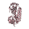
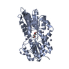
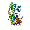
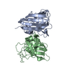
- PDB-6hbm: Crystal structure of MSMEG_1712 from Mycobacterium smegmatis in c... -
+ Open data

- Basic information
| Entry | Database: PDB / ID: 6hbm | |||||||||
|---|---|---|---|---|---|---|---|---|---|---|
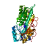
| Title | Crystal structure of MSMEG_1712 from Mycobacterium smegmatis in complex with alpha-L-arabinofuranose | |||||||||
 Components Components | ABC transporter periplasmic-binding protein YtfQ | |||||||||
 Keywords Keywords | SUGAR BINDING PROTEIN / periplasmic binding protein | |||||||||
| Function / homology |  Function and homology information Function and homology information | |||||||||
| Biological species |  Mycobacterium smegmatis (bacteria) Mycobacterium smegmatis (bacteria) | |||||||||
| Method |  X-RAY DIFFRACTION / SYNCHROTRON / MOLECULAR REPLACEMENT / Resolution: 2.76 Å X-RAY DIFFRACTION / SYNCHROTRON / MOLECULAR REPLACEMENT / Resolution: 2.76 Å | |||||||||
 Authors Authors | Li, M. / Mueller, C. / Einsle, O. / Jessen-Trefzer, C. | |||||||||
 Citation Citation | Journal: Cell Chem Biol / Year: 2019 Title: Detection and Characterization of a Mycobacterial L-Arabinofuranose ABC Transporter Identified with a Rapid Lipoproteomics Protocol. Authors: Li, M. / Muller, C. / Frohlich, K. / Gorka, O. / Zhang, L. / Gross, O. / Schilling, O. / Einsle, O. / Jessen-Trefzer, C. | |||||||||
| History |
|
- Structure visualization
| Structure viewer | Molecule: MolmilJmol/JSmol |
|---|
- Downloads & links
-Download
| PDBx/mmCIF format | 6hbm.cif.gz | 226.1 KB | Display | PDBx/mmCIF format |
|---|---|---|---|---|
| PDB format | pdb6hbm.ent.gz | 181.9 KB | Display | PDB format |
| PDBx/mmJSON format | 6hbm.json.gz | Tree view | PDBx/mmJSON format | |
| Others |  Other downloads Other downloads |
-Validation report
| Arichive directory | https://data.pdbj.org/pub/pdb/validation_reports/hb/6hbmftp://data.pdbj.org/pub/pdb/validation_reports/hb/6hbm | HTTPS FTP |
|---|
-Related structure data
| Related structure data |  6hb0SC  6hbdC  6hyhC S: Starting model for refinement C: citing same article ( |
|---|---|
| Similar structure data |


-Links
 PDBj
PDBj


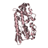
- Assembly
| Deposited unit | 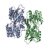
| ||||||||
|---|---|---|---|---|---|---|---|---|---|
| 1 | 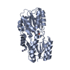
| ||||||||
| 2 | 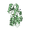
| ||||||||
| Unit cell |
| ||||||||
| Components on special symmetry positions |
|
-Components
| #1: Protein | Mass: 33830.684 Da / Num. of mol.: 2 Source method: isolated from a genetically manipulated source Source: (gene. exp.) Mycobacterium smegmatis (strain ATCC 700084 / mc(2)155) (bacteria)Strain: ATCC 700084 / mc(2)155 / Gene: MSMEG_1712 / Production host: #2: Chemical | ChemComp-ZN /   Mass: 65.409 Da / Num. of mol.: 12 / Source method: obtained synthetically / Formula: Zn Mass: 65.409 Da / Num. of mol.: 12 / Source method: obtained synthetically / Formula: Zn#3: Sugar | 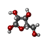  Type: L-saccharide, alpha linking / Mass: 150.130 Da / Num. of mol.: 2 Type: L-saccharide, alpha linking / Mass: 150.130 Da / Num. of mol.: 2Source method: isolated from a genetically manipulated source Formula: C5H10O5 #4: Water | ChemComp-HOH / |  Mass: 18.015 Da / Num. of mol.: 16 / Source method: isolated from a natural source / Formula: H2O Mass: 18.015 Da / Num. of mol.: 16 / Source method: isolated from a natural source / Formula: H2O |
|---|
-Experimental details
-Experiment
| Experiment | Method: X-RAY DIFFRACTION / Number of used crystals: 1 |
|---|
- Sample preparation
| Crystal | Density Matthews: 3.1 Å3/Da / Density % sol: 60.28 % |
|---|---|
| Crystal grow | Temperature: 293 K / Method: vapor diffusion, sitting drop / pH: 8 Details: 0.1 M Tris, pH 8.0 21 % PEG 400 0.325 M NaOAc 0.1 M NaCl |
-Data collection
| Diffraction | Mean temperature: 100 K |
|---|---|
| Diffraction source | Source: SYNCHROTRON / Site: SLS  / Beamline: X06SA / Wavelength: 1 Å / Beamline: X06SA / Wavelength: 1 Å |
| Detector | Type: DECTRIS EIGER X 16M / Detector: PIXEL / Date: Feb 3, 2018 |
| Radiation | Protocol: SINGLE WAVELENGTH / Monochromatic (M) / Laue (L): M / Scattering type: x-ray |
| Radiation wavelength | Wavelength: 1 Å / Relative weight: 1 |
| Reflection | Resolution: 2.75→106.5 Å / Num. obs: 63068 / % possible obs: 92.9 % / Redundancy: 25.7 % / Biso Wilson estimate: 109.79 Å2 / Net I/σ(I): 17.5 |
| Reflection shell | Resolution: 2.75→3.19 Å |
- Processing
| Software |
| ||||||||||||||||||||||||||||||||||||||||||||||||||||||||||||||||||||||||||||||||||||||||||||||||||||||||||||||||||
|---|---|---|---|---|---|---|---|---|---|---|---|---|---|---|---|---|---|---|---|---|---|---|---|---|---|---|---|---|---|---|---|---|---|---|---|---|---|---|---|---|---|---|---|---|---|---|---|---|---|---|---|---|---|---|---|---|---|---|---|---|---|---|---|---|---|---|---|---|---|---|---|---|---|---|---|---|---|---|---|---|---|---|---|---|---|---|---|---|---|---|---|---|---|---|---|---|---|---|---|---|---|---|---|---|---|---|---|---|---|---|---|---|---|---|---|
| Refinement | Method to determine structure: MOLECULAR REPLACEMENT Starting model: 6HB0 Resolution: 2.76→28.95 Å / Cor.coef. Fo:Fc: 0.922 / Cor.coef. Fo:Fc free: 0.861 / Cross valid method: THROUGHOUT / σ(F): 0 / SU Rfree Blow DPI: 0.606
| ||||||||||||||||||||||||||||||||||||||||||||||||||||||||||||||||||||||||||||||||||||||||||||||||||||||||||||||||||
| Displacement parameters | Biso mean: 96.95 Å2
| ||||||||||||||||||||||||||||||||||||||||||||||||||||||||||||||||||||||||||||||||||||||||||||||||||||||||||||||||||
| Refine analyze | Luzzati coordinate error obs: 0.49 Å | ||||||||||||||||||||||||||||||||||||||||||||||||||||||||||||||||||||||||||||||||||||||||||||||||||||||||||||||||||
| Refinement step | Cycle: 1 / Resolution: 2.76→28.95 Å
| ||||||||||||||||||||||||||||||||||||||||||||||||||||||||||||||||||||||||||||||||||||||||||||||||||||||||||||||||||
| Refine LS restraints |
| ||||||||||||||||||||||||||||||||||||||||||||||||||||||||||||||||||||||||||||||||||||||||||||||||||||||||||||||||||
| LS refinement shell | Resolution: 2.76→3.14 Å / Total num. of bins used: 26
|
