| Entry | Database: PDB / ID: 5lpg
|
|---|
| Title | Structure of NUDT15 in complex with 6-thio-GMP |
|---|
 Components Components | Probable 8-oxo-dGTP diphosphatase NUDT15 |
|---|
 Keywords Keywords | HYDROLASE / MTH2 / DNA repair enzyme / Cancer |
|---|
| Function / homology |  Function and homology information Function and homology information
nucleoside phosphate catabolic process / prenyl-diphosphate phosphatase / isoprenoid diphosphate phosphatase activity / geranyl diphosphate phosphohydrolase / purine nucleotide catabolic process / nucleotide diphosphatase / nucleobase-containing small molecule metabolic process / nucleoside triphosphate diphosphatase activity / 8-oxo-dGDP phosphatase activity / dGTP catabolic process ...nucleoside phosphate catabolic process / prenyl-diphosphate phosphatase / isoprenoid diphosphate phosphatase activity / geranyl diphosphate phosphohydrolase / purine nucleotide catabolic process / nucleotide diphosphatase / nucleobase-containing small molecule metabolic process / nucleoside triphosphate diphosphatase activity / 8-oxo-dGDP phosphatase activity / dGTP catabolic process / 8-oxo-7,8-dihydrodeoxyguanosine triphosphate pyrophosphatase activity / 8-oxo-7,8-dihydroguanosine triphosphate pyrophosphatase activity / DNA protection / Phosphate bond hydrolysis by NUDT proteins / Azathioprine ADME / xenobiotic catabolic process / regulation of proteasomal protein catabolic process / response to reactive oxygen species / mitotic cell cycle / metal ion binding / cytosolSimilarity search - Function Nucleoside Triphosphate Pyrophosphohydrolase / Nucleoside Triphosphate Pyrophosphohydrolase / NUDIX domain / Nudix hydrolase domain profile. / NUDIX hydrolase domain / NUDIX hydrolase-like domain superfamily / Alpha-Beta Complex / Alpha BetaSimilarity search - Domain/homology |
|---|
| Biological species |  Homo sapiens (human) Homo sapiens (human) |
|---|
| Method |  X-RAY DIFFRACTION / SYNCHROTRON / MOLECULAR REPLACEMENT / Resolution: 1.7 Å X-RAY DIFFRACTION / SYNCHROTRON / MOLECULAR REPLACEMENT / Resolution: 1.7 Å |
|---|
 Authors Authors | Masuyer, G. / Carter, M. / Rehling, D. / Stenmark, P. / Helleday, T. / Jemth, A.-S. / Valerie, N.C.K. / Homan, E. / Herr, P. / Bevc, L. ...Masuyer, G. / Carter, M. / Rehling, D. / Stenmark, P. / Helleday, T. / Jemth, A.-S. / Valerie, N.C.K. / Homan, E. / Herr, P. / Bevc, L. / Page, B.D.G. / Hagenkort, A. |
|---|
| Funding support |  Sweden, 1items Sweden, 1items | Organization | Grant number | Country |
|---|
| | Sweden |
|
|---|
 Citation Citation | Journal: Cancer Res. / Year: 2016
Title: NUDT15 Hydrolyzes 6-Thio-DeoxyGTP to Mediate the Anticancer Efficacy of 6-Thioguanine.
Authors: Valerie, N.C. / Hagenkort, A. / Page, B.D. / Masuyer, G. / Rehling, D. / Carter, M. / Bevc, L. / Herr, P. / Homan, E. / Sheppard, N.G. / Stenmark, P. / Jemth, A.S. / Helleday, T. |
|---|
| History | | Deposition | Aug 12, 2016 | Deposition site: PDBE / Processing site: PDBE |
|---|
| Revision 1.0 | Aug 24, 2016 | Provider: repository / Type: Initial release |
|---|
| Revision 1.1 | Sep 14, 2016 | Group: Database references |
|---|
| Revision 1.2 | Sep 28, 2016 | Group: Database references |
|---|
| Revision 1.3 | Jan 10, 2024 | Group: Data collection / Database references ...Data collection / Database references / Derived calculations / Refinement description
Category: chem_comp_atom / chem_comp_bond ...chem_comp_atom / chem_comp_bond / database_2 / pdbx_initial_refinement_model / pdbx_struct_conn_angle / struct_conn
Item: _database_2.pdbx_DOI / _database_2.pdbx_database_accession ..._database_2.pdbx_DOI / _database_2.pdbx_database_accession / _pdbx_struct_conn_angle.ptnr1_auth_asym_id / _pdbx_struct_conn_angle.ptnr1_auth_comp_id / _pdbx_struct_conn_angle.ptnr1_auth_seq_id / _pdbx_struct_conn_angle.ptnr1_label_asym_id / _pdbx_struct_conn_angle.ptnr1_label_atom_id / _pdbx_struct_conn_angle.ptnr1_label_comp_id / _pdbx_struct_conn_angle.ptnr1_label_seq_id / _pdbx_struct_conn_angle.ptnr2_auth_seq_id / _pdbx_struct_conn_angle.ptnr2_label_asym_id / _pdbx_struct_conn_angle.ptnr3_auth_asym_id / _pdbx_struct_conn_angle.ptnr3_auth_comp_id / _pdbx_struct_conn_angle.ptnr3_auth_seq_id / _pdbx_struct_conn_angle.ptnr3_label_asym_id / _pdbx_struct_conn_angle.ptnr3_label_atom_id / _pdbx_struct_conn_angle.ptnr3_label_comp_id / _pdbx_struct_conn_angle.ptnr3_label_seq_id / _pdbx_struct_conn_angle.value / _struct_conn.pdbx_dist_value / _struct_conn.ptnr1_auth_asym_id / _struct_conn.ptnr1_auth_comp_id / _struct_conn.ptnr1_auth_seq_id / _struct_conn.ptnr1_label_asym_id / _struct_conn.ptnr1_label_atom_id / _struct_conn.ptnr1_label_comp_id / _struct_conn.ptnr1_label_seq_id / _struct_conn.ptnr2_auth_asym_id / _struct_conn.ptnr2_auth_comp_id / _struct_conn.ptnr2_auth_seq_id / _struct_conn.ptnr2_label_asym_id / _struct_conn.ptnr2_label_atom_id / _struct_conn.ptnr2_label_comp_id |
|---|
|
|---|
 Movie
Movie Controller


 Open data
 Basic information
 Structure visualization
 Downloads & links
 Other downloads
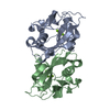


 PDBj
PDBj


 Assembly


 Mass: 24.305 Da / Num. of mol.: 5 / Source method: obtained synthetically / Formula: Mg
 Mass: 379.286 Da / Num. of mol.: 1 / Source method: obtained synthetically / Formula: C10H14N5O7PS
 Mass: 18.015 Da / Num. of mol.: 156 / Source method: isolated from a natural source / Formula: H2O
 Sample preparation
 / Beamline: ID29 / Wavelength: 1.072 Å
 Processing