 Movie
Movie Controller

[English] 日本語
 Yorodumi
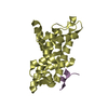
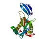
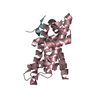
- PDB-7azn: Structure of mouse AsterC (GramD1c) with a new cholesterol-derive... -
+ Open data

- Basic information
| Entry | Database: PDB / ID: 7azn | ||||||
|---|---|---|---|---|---|---|---|
| Title | Structure of mouse AsterC (GramD1c) with a new cholesterol-derived compound | ||||||
 Components Components | Protein Aster-C | ||||||
 Keywords Keywords | LIPID TRANSPORT / Non vesicular cholesterol transport / lipid-binding / endoplasmic reticulum / membrane contact sites | ||||||
| Function / homology |  Function and homology information Function and homology informationintracellular sterol transport / endoplasmic reticulum-plasma membrane contact site / cellular response to cholesterol / cholesterol transfer activity / cholesterol binding / endoplasmic reticulum membrane / plasma membrane Similarity search - Function | ||||||
| Biological species |  | ||||||
| Method |  X-RAY DIFFRACTION / SYNCHROTRON / MOLECULAR REPLACEMENT / Resolution: 2.09 Å X-RAY DIFFRACTION / SYNCHROTRON / MOLECULAR REPLACEMENT / Resolution: 2.09 Å | ||||||
 Authors Authors | Romartinez-Alonso, B. / Sirvydis, K. / Kim, Y. / Xiao, X. / Jung, M. / Tontonoz, P. / Schwabe, J. | ||||||
| Funding support |  France, 1items France, 1items
| ||||||
 Citation Citation | Journal: Proc.Natl.Acad.Sci.USA / Year: 2021 Title: Selective Aster inhibitors distinguish vesicular and nonvesicular sterol transport mechanisms. Authors: Xiao, X. / Kim, Y. / Romartinez-Alonso, B. / Sirvydis, K. / Ory, D.S. / Schwabe, J.W.R. / Jung, M.E. / Tontonoz, P. | ||||||
| History |
|
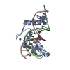
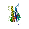
- Structure visualization
| Structure viewer | Molecule: MolmilJmol/JSmol |
|---|
- Downloads & links
-Download
| PDBx/mmCIF format | 7azn.cif.gz | 54.3 KB | Display | PDBx/mmCIF format |
|---|---|---|---|---|
| PDB format | pdb7azn.ent.gz | 36.8 KB | Display | PDB format |
| PDBx/mmJSON format | 7azn.json.gz | Tree view | PDBx/mmJSON format | |
| Others |  Other downloads Other downloads |
-Validation report
| Arichive directory | https://data.pdbj.org/pub/pdb/validation_reports/az/7aznftp://data.pdbj.org/pub/pdb/validation_reports/az/7azn | HTTPS FTP |
|---|
-Related structure data
| Related structure data | 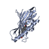 6gqfS S: Starting model for refinement |
|---|---|
| Similar structure data |


-Links
 PDBj
PDBj


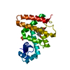
- Assembly
| Deposited unit | 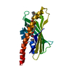
| ||||||||
|---|---|---|---|---|---|---|---|---|---|
| 1 |
| ||||||||
| Unit cell |
|
-Components
| #1: Protein | Mass: 20833.688 Da / Num. of mol.: 1 Source method: isolated from a genetically manipulated source Source: (gene. exp.)  | ||||||
|---|---|---|---|---|---|---|---|
| #2: Chemical | ChemComp-YK8 / 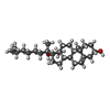  Mass: 416.680 Da / Num. of mol.: 1 / Source method: obtained synthetically / Formula: C28H48O2 / Feature type: SUBJECT OF INVESTIGATION Mass: 416.680 Da / Num. of mol.: 1 / Source method: obtained synthetically / Formula: C28H48O2 / Feature type: SUBJECT OF INVESTIGATION | ||||||
| #3: Chemical | ChemComp-EOH /   Mass: 46.068 Da / Num. of mol.: 6 / Source method: obtained synthetically / Formula: C2H6O Mass: 46.068 Da / Num. of mol.: 6 / Source method: obtained synthetically / Formula: C2H6O#4: Chemical | ChemComp-GOL / |   Mass: 92.094 Da / Num. of mol.: 1 / Source method: obtained synthetically / Formula: C3H8O3 Mass: 92.094 Da / Num. of mol.: 1 / Source method: obtained synthetically / Formula: C3H8O3#5: Water | ChemComp-HOH / |  Mass: 18.015 Da / Num. of mol.: 50 / Source method: isolated from a natural source / Formula: H2O Mass: 18.015 Da / Num. of mol.: 50 / Source method: isolated from a natural source / Formula: H2OHas ligand of interest | Y | |
-Experimental details
-Experiment
| Experiment | Method: X-RAY DIFFRACTION / Number of used crystals: 1 |
|---|
- Sample preparation
| Crystal | Density Matthews: 2.32 Å3/Da / Density % sol: 47 % / Description: A single 100-micrometres rectangular prism. |
|---|---|
| Crystal grow | Temperature: 291 K / Method: vapor diffusion, sitting drop / pH: 4 / Details: 0.1 M SPG buffer pH 4 25 % PEG 1500 |
-Data collection
| Diffraction | Mean temperature: 293 K / Serial crystal experiment: N |
|---|---|
| Diffraction source | Source: SYNCHROTRON / Site: Diamond  / Beamline: I24 / Wavelength: 0.976 Å / Beamline: I24 / Wavelength: 0.976 Å |
| Detector | Type: DECTRIS PILATUS3 6M / Detector: PIXEL / Date: Dec 15, 2019 / Details: Mirrors |
| Radiation | Monochromator: Double crystal monochromator 28.900 m / Protocol: SINGLE WAVELENGTH / Monochromatic (M) / Laue (L): M / Scattering type: x-ray |
| Radiation wavelength | Wavelength: 0.976 Å / Relative weight: 1 |
| Reflection | Resolution: 2.09→26.77 Å / Num. obs: 11465 / % possible obs: 97.9 % / Redundancy: 3.2 % / CC1/2: 0.994 / Rmerge(I) obs: 0.06 / Rpim(I) all: 0.057 / Rrim(I) all: 0.083 / Χ2: 0.69 / Net I/σ(I): 7.1 |
| Reflection shell | Resolution: 2.09→2.15 Å / Redundancy: 2.7 % / Rmerge(I) obs: 0.252 / Mean I/σ(I) obs: 2.9 / Num. unique obs: 808 / CC1/2: 0.944 / Rpim(I) all: 0.239 / Rrim(I) all: 0.347 / Χ2: 0.65 / % possible all: 84.2 |
- Processing
| Software |
| |||||||||||||||||||||||||||||||||||
|---|---|---|---|---|---|---|---|---|---|---|---|---|---|---|---|---|---|---|---|---|---|---|---|---|---|---|---|---|---|---|---|---|---|---|---|---|
| Refinement | Method to determine structure: MOLECULAR REPLACEMENT Starting model: 6gqf Resolution: 2.09→26.77 Å / SU ML: 0.27 / Cross valid method: THROUGHOUT / σ(F): 1.35 / Phase error: 35.67 / Stereochemistry target values: ML
| |||||||||||||||||||||||||||||||||||
| Solvent computation | Shrinkage radii: 0.9 Å / VDW probe radii: 1.11 Å / Solvent model: FLAT BULK SOLVENT MODEL | |||||||||||||||||||||||||||||||||||
| Displacement parameters | Biso max: 92.05 Å2 / Biso mean: 41.7578 Å2 / Biso min: 16.36 Å2 | |||||||||||||||||||||||||||||||||||
| Refinement step | Cycle: final / Resolution: 2.09→26.77 Å
| |||||||||||||||||||||||||||||||||||
| LS refinement shell | Refine-ID: X-RAY DIFFRACTION / Rfactor Rfree error: 0 / Total num. of bins used: 4
|
