 Movie
Movie Controller

[English] 日本語
 Yorodumi
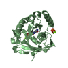
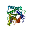
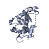
- PDB-7ax7: Crystal structure of the Xyl-CE4 domain of a multidomain xylanase... -
+ Open data

- Basic information
| Entry | Database: PDB / ID: 7ax7 | ||||||
|---|---|---|---|---|---|---|---|
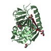
| Title | Crystal structure of the Xyl-CE4 domain of a multidomain xylanase from the hindgut metagenome of Trinervitermes trinervoides | ||||||
 Components Components | Endo-1,4-beta-xylanase | ||||||
 Keywords Keywords | METAL BINDING PROTEIN / Xyl-CE4 uses Co2+ to cleave acetyl groups from acetylated xylan molecules. As part of a multidomain assembly / CE4 functions independently of other domain components. | ||||||
| Function / homology |  Function and homology information Function and homology informationhydrolase activity, acting on carbon-nitrogen (but not peptide) bonds / endo-1,4-beta-xylanase / endo-1,4-beta-xylanase activity / xylan catabolic process / carbohydrate binding / metal ion binding Similarity search - Function | ||||||
| Biological species |  uncultured bacterium (environmental samples) uncultured bacterium (environmental samples) | ||||||
| Method |  X-RAY DIFFRACTION / MOLECULAR REPLACEMENT / Resolution: 2.05000181627 Å X-RAY DIFFRACTION / MOLECULAR REPLACEMENT / Resolution: 2.05000181627 Å | ||||||
 Authors Authors | Anye, V. / Schubert, W.D. | ||||||
| Funding support |  South Africa, 1items South Africa, 1items
| ||||||
 Citation Citation | Journal: Plos One / Year: 2022 Title: Structural and biophysical characterization of the multidomain xylanase Xyl. Authors: Anye, V. / Kruger, R.F. / Schubert, W.D. | ||||||
| History |
|
- Structure visualization
| Structure viewer | Molecule: MolmilJmol/JSmol |
|---|
- Downloads & links
-Download
| PDBx/mmCIF format | 7ax7.cif.gz | 73.2 KB | Display | PDBx/mmCIF format |
|---|---|---|---|---|
| PDB format | pdb7ax7.ent.gz | 42.3 KB | Display | PDB format |
| PDBx/mmJSON format | 7ax7.json.gz | Tree view | PDBx/mmJSON format | |
| Others |  Other downloads Other downloads |
-Validation report
| Arichive directory | https://data.pdbj.org/pub/pdb/validation_reports/ax/7ax7ftp://data.pdbj.org/pub/pdb/validation_reports/ax/7ax7 | HTTPS FTP |
|---|
-Related structure data
| Related structure data |  7ay3C  7aypC 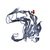 7zszC  2c71S S: Starting model for refinement C: citing same article ( |
|---|---|
| Similar structure data |


-Links
 PDBj
PDBj


- Assembly
| Deposited unit | 
| ||||||||||||
|---|---|---|---|---|---|---|---|---|---|---|---|---|---|
| 1 |
| ||||||||||||
| Unit cell |
|
-Components
| #1: Protein | Mass: 23812.477 Da / Num. of mol.: 1 Source method: isolated from a genetically manipulated source Source: (gene. exp.) uncultured bacterium (environmental samples)Production host: |
|---|---|
| #2: Chemical | ChemComp-CO /   Mass: 58.933 Da / Num. of mol.: 1 / Source method: isolated from a natural source / Formula: Co Mass: 58.933 Da / Num. of mol.: 1 / Source method: isolated from a natural source / Formula: Co |
| #3: Chemical | ChemComp-ACT /   Mass: 59.044 Da / Num. of mol.: 1 / Source method: obtained synthetically / Formula: C2H3O2 Mass: 59.044 Da / Num. of mol.: 1 / Source method: obtained synthetically / Formula: C2H3O2 |
| #4: Water | ChemComp-HOH /  Mass: 18.015 Da / Num. of mol.: 247 / Source method: isolated from a natural source / Formula: H2O Mass: 18.015 Da / Num. of mol.: 247 / Source method: isolated from a natural source / Formula: H2O |
| Has ligand of interest | N |
-Experimental details
-Experiment
| Experiment | Method: X-RAY DIFFRACTION / Number of used crystals: 1 |
|---|
- Sample preparation
| Crystal | Density Matthews: 2.17 Å3/Da / Density % sol: 43.2 % |
|---|---|
| Crystal grow | Temperature: 292.2 K / Method: vapor diffusion, sitting drop / pH: 7.5 Details: 10 mg/mL in 20 mM Tris-HCl pH 7.5, 10 mM NaCl, 8 % (w/v) PEG 4000 |
-Data collection
| Diffraction | Mean temperature: 100 K / Serial crystal experiment: N |
|---|---|
| Diffraction source | Source: ROTATING ANODE / Type: RIGAKU / Wavelength: 1.54 Å |
| Detector | Type: RIGAKU SATURN 944 / Detector: CCD / Date: Apr 9, 2017 |
| Radiation | Protocol: SINGLE WAVELENGTH / Monochromatic (M) / Laue (L): M / Scattering type: x-ray |
| Radiation wavelength | Wavelength: 1.54 Å / Relative weight: 1 |
| Reflection | Resolution: 1.93→23.07 Å / Num. obs: 13512 / % possible obs: 99 % / Redundancy: 2 % / Biso Wilson estimate: 13.713555817 Å2 / CC1/2: 0.999 / Rpim(I) all: 0.019 / Net I/av σ(I): 18 / Net I/σ(I): 2.74 |
| Reflection shell | Resolution: 2.05→2.12 Å / Num. unique obs: 722 / CC1/2: 0.991 / % possible all: 96.9 |
- Processing
| Software |
| ||||||||||||||||||||||||||||||||||||||||||
|---|---|---|---|---|---|---|---|---|---|---|---|---|---|---|---|---|---|---|---|---|---|---|---|---|---|---|---|---|---|---|---|---|---|---|---|---|---|---|---|---|---|---|---|
| Refinement | Method to determine structure: MOLECULAR REPLACEMENT Starting model: 2C71 Resolution: 2.05000181627→19.5413775456 Å / SU ML: 0.250769489372 / Cross valid method: FREE R-VALUE / σ(F): 1.4376150803 / Phase error: 25.4581162604 Stereochemistry target values: GeoStd + Monomer Library + CDL v1.2
| ||||||||||||||||||||||||||||||||||||||||||
| Solvent computation | Shrinkage radii: 0.9 Å / VDW probe radii: 1.11 Å / Solvent model: FLAT BULK SOLVENT MODEL | ||||||||||||||||||||||||||||||||||||||||||
| Displacement parameters | Biso mean: 14.1404325504 Å2 | ||||||||||||||||||||||||||||||||||||||||||
| Refinement step | Cycle: LAST / Resolution: 2.05000181627→19.5413775456 Å
| ||||||||||||||||||||||||||||||||||||||||||
| Refine LS restraints |
| ||||||||||||||||||||||||||||||||||||||||||
| LS refinement shell |
|
