 Movie
Movie Controller

[English] 日本語
 Yorodumi
- PDB-7ayp: Structure of a GH11 domain refined from the X-ray diffraction dat... -
+ Open data

- Basic information
| Entry | Database: PDB / ID: 7ayp | ||||||
|---|---|---|---|---|---|---|---|
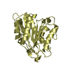
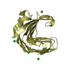
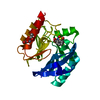
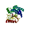
| Title | Structure of a GH11 domain refined from the X-ray diffraction data of a GH11-CBM36-1 crystal. | ||||||
 Components Components | Endo-1,4-beta-xylanase | ||||||
 Keywords Keywords | HYDROLASE / Multidomain protein / carbohydrate binding domains / GH11 / interdomain interactions / carbohydrate esterases / Isothermal titration calorimetry | ||||||
| Function / homology |  Function and homology information Function and homology informationhydrolase activity, acting on carbon-nitrogen (but not peptide) bonds / endo-1,4-beta-xylanase / endo-1,4-beta-xylanase activity / xylan catabolic process / carbohydrate binding / metal ion binding Similarity search - Function | ||||||
| Biological species |  uncultured bacterium (environmental samples) uncultured bacterium (environmental samples) | ||||||
| Method |  X-RAY DIFFRACTION / SYNCHROTRON / MOLECULAR REPLACEMENT / Resolution: 1.70001286876 Å X-RAY DIFFRACTION / SYNCHROTRON / MOLECULAR REPLACEMENT / Resolution: 1.70001286876 Å | ||||||
 Authors Authors | Anye, V. / Schubert, W.D. | ||||||
| Funding support |  South Africa, 1items South Africa, 1items
| ||||||
 Citation Citation | Journal: Plos One / Year: 2022 Title: Structural and biophysical characterization of the multidomain xylanase Xyl. Authors: Anye, V. / Kruger, R.F. / Schubert, W.D. | ||||||
| History |
|
- Structure visualization
| Structure viewer | Molecule: MolmilJmol/JSmol |
|---|
- Downloads & links
-Download
| PDBx/mmCIF format | 7ayp.cif.gz | 72.7 KB | Display | PDBx/mmCIF format |
|---|---|---|---|---|
| PDB format | pdb7ayp.ent.gz | 41.1 KB | Display | PDB format |
| PDBx/mmJSON format | 7ayp.json.gz | Tree view | PDBx/mmJSON format | |
| Others |  Other downloads Other downloads |
-Validation report
| Arichive directory | https://data.pdbj.org/pub/pdb/validation_reports/ay/7aypftp://data.pdbj.org/pub/pdb/validation_reports/ay/7ayp | HTTPS FTP |
|---|
-Related structure data
| Related structure data |  7ax7C  7ay3C  7zszC  2dcjS S: Starting model for refinement C: citing same article ( |
|---|---|
| Similar structure data |


-Links
 PDBj
PDBj

- Assembly
| Deposited unit | 
| ||||||||||||
|---|---|---|---|---|---|---|---|---|---|---|---|---|---|
| 1 |
| ||||||||||||
| Unit cell |
| ||||||||||||
| Components on special symmetry positions |
|
-Components
| #1: Protein | Mass: 46589.574 Da / Num. of mol.: 1 Source method: isolated from a genetically manipulated source Details: GH11-CBM36-1 Source: (gene. exp.) uncultured bacterium (environmental samples)Production host: |
|---|---|
| #2: Water | ChemComp-HOH /  Mass: 18.015 Da / Num. of mol.: 136 / Source method: isolated from a natural source / Formula: H2O Mass: 18.015 Da / Num. of mol.: 136 / Source method: isolated from a natural source / Formula: H2O |
-Experimental details
-Experiment
| Experiment | Method: X-RAY DIFFRACTION / Number of used crystals: 1 |
|---|
- Sample preparation
| Crystal | Density Matthews: 1.97 Å3/Da / Density % sol: 37.48 % |
|---|---|
| Crystal grow | Temperature: 293.16 K / Method: vapor diffusion, hanging drop / pH: 4.8 / Details: Na acetate, (NH4)2SO4 |
-Data collection
| Diffraction | Mean temperature: 100 K / Serial crystal experiment: N |
|---|---|
| Diffraction source | Source: SYNCHROTRON / Site: ESRF  / Beamline: ID29 / Wavelength: 0.65 Å / Beamline: ID29 / Wavelength: 0.65 Å |
| Detector | Type: DECTRIS PILATUS 6M-F / Detector: PIXEL / Date: Dec 14, 2016 |
| Radiation | Protocol: SINGLE WAVELENGTH / Monochromatic (M) / Laue (L): M / Scattering type: x-ray |
| Radiation wavelength | Wavelength: 0.65 Å / Relative weight: 1 |
| Reflection | Resolution: 1.47→93.69 Å / Num. obs: 65364 / % possible obs: 99.9 % / Redundancy: 1 % / Biso Wilson estimate: 14.6903258564 Å2 / CC1/2: 0.999 / Net I/σ(I): 15.81 |
| Reflection shell | Resolution: 1.47→3.9 Å / Num. unique obs: 3225 / CC1/2: 0.814 / % possible all: 99 |
- Processing
| Software | Name: PHENIX / Version: 1.12_2829 / Classification: refinement | |||||||||||||||||||||||||||||||||||||||||||||||||||||||||||||||||||||||||||||||||||||||||||||||||||||||||
|---|---|---|---|---|---|---|---|---|---|---|---|---|---|---|---|---|---|---|---|---|---|---|---|---|---|---|---|---|---|---|---|---|---|---|---|---|---|---|---|---|---|---|---|---|---|---|---|---|---|---|---|---|---|---|---|---|---|---|---|---|---|---|---|---|---|---|---|---|---|---|---|---|---|---|---|---|---|---|---|---|---|---|---|---|---|---|---|---|---|---|---|---|---|---|---|---|---|---|---|---|---|---|---|---|---|---|
| Refinement | Method to determine structure: MOLECULAR REPLACEMENT Starting model: 2DCJ Resolution: 1.70001286876→54.108 Å / SU ML: 0.143195300177 / Cross valid method: FREE R-VALUE / σ(F): 1.33571280734 / Phase error: 17.930989269 Stereochemistry target values: GeoStd + Monomer Library + CDL v1.2
| |||||||||||||||||||||||||||||||||||||||||||||||||||||||||||||||||||||||||||||||||||||||||||||||||||||||||
| Solvent computation | Shrinkage radii: 0.9 Å / VDW probe radii: 1.11 Å / Solvent model: FLAT BULK SOLVENT MODEL | |||||||||||||||||||||||||||||||||||||||||||||||||||||||||||||||||||||||||||||||||||||||||||||||||||||||||
| Displacement parameters | Biso mean: 16.9406706224 Å2 | |||||||||||||||||||||||||||||||||||||||||||||||||||||||||||||||||||||||||||||||||||||||||||||||||||||||||
| Refinement step | Cycle: LAST / Resolution: 1.70001286876→54.108 Å
| |||||||||||||||||||||||||||||||||||||||||||||||||||||||||||||||||||||||||||||||||||||||||||||||||||||||||
| Refine LS restraints |
| |||||||||||||||||||||||||||||||||||||||||||||||||||||||||||||||||||||||||||||||||||||||||||||||||||||||||
| LS refinement shell |
|
