 Movie
Movie Controller

[English] 日本語
 Yorodumi
- PDB-3cn7: Crystal Structure Analysis of the Carboxylesterase PA3859 from Ps... -
+ Open data

- Basic information
| Entry | Database: PDB / ID: 3cn7 | ||||||
|---|---|---|---|---|---|---|---|
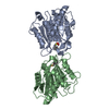
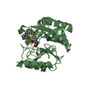
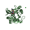
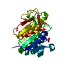
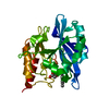
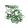
| Title | Crystal Structure Analysis of the Carboxylesterase PA3859 from Pseudomonas aeruginosa PAO1- MONOCLINIC CRYSTAL FORM | ||||||
 Components Components | Carboxylesterase | ||||||
 Keywords Keywords | HYDROLASE / alpha/beta hydrolase fold super-family | ||||||
| Function / homology |  Function and homology information Function and homology information | ||||||
| Biological species |   Pseudomonas aeruginosa (bacteria) Pseudomonas aeruginosa (bacteria) | ||||||
| Method |  X-RAY DIFFRACTION / SYNCHROTRON / MOLECULAR REPLACEMENT / Resolution: 2.99 Å X-RAY DIFFRACTION / SYNCHROTRON / MOLECULAR REPLACEMENT / Resolution: 2.99 Å | ||||||
 Authors Authors | Pesaresi, A. / Lamba, D. | ||||||
 Citation Citation | Journal: Biochimie / Year: 2010 Title: Insights into the fatty acid chain length specificity of the carboxylesterase PA3859 from Pseudomonas aeruginosa: A combined structural, biochemical and computational study. Authors: Pesaresi, A. / Lamba, D. #1: Journal: CURR.MICROBIOL. / Year: 2005 Title: Isolation, Characterization, and Heterologous Expression of a Carboxylesterase of Pseudomonas aeruginosa PAO1 Authors: Pesaresi, A. / Devescovi, G. / Lamba, D. / Venturi, V. / Degrassi, G. #2: Journal: BIOCHEM.BIOPHYS.ACTA PROTEINS & PROTEOMICS / Year: 2005 Title: Crystallization, X-ray Diffraction Analysis and Phasing of Carboxylesterase PA3859 from Pseudomonas aeruginosa Authors: Pesaresi, A. / Lamba, D. | ||||||
| History |
|
- Structure visualization
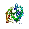
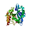
| Structure viewer | Molecule: MolmilJmol/JSmol |
|---|
- Downloads & links
-Download
| PDBx/mmCIF format | 3cn7.cif.gz | 172.1 KB | Display | PDBx/mmCIF format |
|---|---|---|---|---|
| PDB format | pdb3cn7.ent.gz | 137.4 KB | Display | PDB format |
| PDBx/mmJSON format | 3cn7.json.gz | Tree view | PDBx/mmJSON format | |
| Others |  Other downloads Other downloads |
-Validation report
| Arichive directory | https://data.pdbj.org/pub/pdb/validation_reports/cn/3cn7ftp://data.pdbj.org/pub/pdb/validation_reports/cn/3cn7 | HTTPS FTP |
|---|
-Related structure data
| Related structure data |  3cn9C 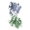 1auoS S: Starting model for refinement C: citing same article ( |
|---|---|
| Similar structure data |


-Links
 PDBj
PDBj- Assembly
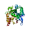
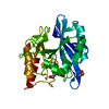
| Deposited unit | 
| ||||||||
|---|---|---|---|---|---|---|---|---|---|
| 1 | 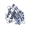
| ||||||||
| 2 | 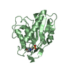
| ||||||||
| 3 | 
| ||||||||
| 4 | 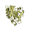
| ||||||||
| Unit cell |
|
-Components
| #1: Protein | Mass: 24719.916 Da / Num. of mol.: 4 Source method: isolated from a genetically manipulated source Source: (gene. exp.) Pseudomonas aeruginosa (bacteria) / Strain: PAO1 / Gene: PA3859 / Plasmid: pQE-31 / Production host: #2: Chemical |   Mass: 195.237 Da / Num. of mol.: 2 / Source method: obtained synthetically / Formula: C6H13NO4S / Comment: pH buffer*YM Mass: 195.237 Da / Num. of mol.: 2 / Source method: obtained synthetically / Formula: C6H13NO4S / Comment: pH buffer*YM#3: Water | ChemComp-HOH / |  Mass: 18.015 Da / Num. of mol.: 94 / Source method: isolated from a natural source / Formula: H2O Mass: 18.015 Da / Num. of mol.: 94 / Source method: isolated from a natural source / Formula: H2OHas protein modification | Y | |
|---|
-Experimental details
-Experiment
| Experiment | Method: X-RAY DIFFRACTION / Number of used crystals: 1 |
|---|
- Sample preparation
| Crystal | Density Matthews: 2.39 Å3/Da / Density % sol: 48.51 % |
|---|---|
| Crystal grow | Temperature: 293 K / pH: 6.5 Details: 0.200 M AMS, 0.100 M MES, 30% w/v PEGMME-5000, Long needles grew within 3 days, pH 6.5, VAPOR DIFFUSION, HANGING DROP, temperature 293K |
-Data collection
| Diffraction | Mean temperature: 100 K |
|---|---|
| Diffraction source | Source: SYNCHROTRON / Site: ELETTRA  / Beamline: 5.2R / Wavelength: 1.2 / Beamline: 5.2R / Wavelength: 1.2 |
| Detector | Type: MAR CCD 165 mm / Detector: CCD / Date: Jul 31, 2002 / Details: THREE-SEGMENT PT-COATED TOROIDAL MIRRORS |
| Radiation | Monochromator: DOUBLE CRYSTAL (SI111) / Protocol: SINGLE WAVELENGTH / Monochromatic (M) / Laue (L): M / Scattering type: x-ray |
| Radiation wavelength | Wavelength: 1.2 Å / Relative weight: 1 |
| Reflection | Resolution: 2.98→24.3 Å / Num. obs: 18803 / % possible obs: 98.3 % / Observed criterion σ(I): -3 / Redundancy: 2.9 % / Biso Wilson estimate: 56.8 Å2 / Rmerge(I) obs: 0.16 / Rsym value: 0.13 / Net I/σ(I): 3.9 |
| Reflection shell | Resolution: 2.98→3 Å / Redundancy: 2.8 % / Rmerge(I) obs: 0.44 / Mean I/σ(I) obs: 1.7 / Rsym value: 0.36 / % possible all: 98.3 |
- Processing
| Software |
| ||||||||||||||||||||||||||||||||||||||||||||||||||||||||||||||||||||||||||||||||
|---|---|---|---|---|---|---|---|---|---|---|---|---|---|---|---|---|---|---|---|---|---|---|---|---|---|---|---|---|---|---|---|---|---|---|---|---|---|---|---|---|---|---|---|---|---|---|---|---|---|---|---|---|---|---|---|---|---|---|---|---|---|---|---|---|---|---|---|---|---|---|---|---|---|---|---|---|---|---|---|---|---|
| Refinement | Method to determine structure: MOLECULAR REPLACEMENT Starting model: PDB ENTRY 1AUO, CHAIN A Resolution: 2.99→23.82 Å / Rfactor Rfree error: 0.006 / Data cutoff high absF: 550394.57 / Data cutoff low absF: 0 / Isotropic thermal model: OVERALL ANISOTROPIC / Cross valid method: THROUGHOUT / σ(F): 0 / Stereochemistry target values: ENGH & HUBER
| ||||||||||||||||||||||||||||||||||||||||||||||||||||||||||||||||||||||||||||||||
| Solvent computation | Solvent model: FLAT MODEL / Bsol: 21.77 Å2 / ksol: 0.35 e/Å3 | ||||||||||||||||||||||||||||||||||||||||||||||||||||||||||||||||||||||||||||||||
| Displacement parameters | Biso mean: 32 Å2
| ||||||||||||||||||||||||||||||||||||||||||||||||||||||||||||||||||||||||||||||||
| Refine analyze |
| ||||||||||||||||||||||||||||||||||||||||||||||||||||||||||||||||||||||||||||||||
| Refinement step | Cycle: LAST / Resolution: 2.99→23.82 Å
| ||||||||||||||||||||||||||||||||||||||||||||||||||||||||||||||||||||||||||||||||
| Refine LS restraints |
| ||||||||||||||||||||||||||||||||||||||||||||||||||||||||||||||||||||||||||||||||
| Refine LS restraints NCS | NCS model details: RESTRAIN | ||||||||||||||||||||||||||||||||||||||||||||||||||||||||||||||||||||||||||||||||
| LS refinement shell | Resolution: 2.99→3.18 Å / Rfactor Rfree error: 0.022 / Total num. of bins used: 6
| ||||||||||||||||||||||||||||||||||||||||||||||||||||||||||||||||||||||||||||||||
| Xplor file |
|
