- PDB-2c71: The structure of a family 4 acetyl xylan esterase from Clostridiu... -
+
Open data
ID or keywords:
Loading...
-
Basic information
Entry
Database: PDB / ID: 2c71
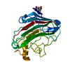
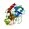
Title
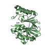
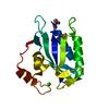
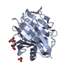
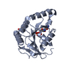
The structure of a family 4 acetyl xylan esterase from Clostridium thermocellum in complex with a magnesium ion.
Components
GLYCOSIDE HYDROLASE, FAMILY 11\:CLOSTRIDIUM CELLULOSOME ENZYME, DOCKERIN TYPE I\:POLYSACCHARIDE
Keywords
HYDROLASE / ACETYL-XYLAN / ESTERASES / METAL-ION
Function / homology
Function and homology information
hydrolase activity, acting on carbon-nitrogen (but not peptide) bonds / endo-1,4-beta-xylanase / endo-1,4-beta-xylanase activity / xylan catabolic process / carbohydrate binding / metal ion binding Similarity search - Function
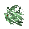
Glycoside hydrolase/deacetylase / NodB homology domain profile. / NodB homology domain / Polysaccharide deacetylase / Cellulose binding, type IV / Cellulose Binding Domain Type IV / Carbohydrate binding module (family 6) / Clostridium cellulosome enzymes repeated domain signature. / CBM6 (carbohydrate binding type-6) domain profile. / Carbohydrate binding module family 6 ...Glycoside hydrolase/deacetylase / NodB homology domain profile. / NodB homology domain / Polysaccharide deacetylase / Cellulose binding, type IV / Cellulose Binding Domain Type IV / Carbohydrate binding module (family 6) / Clostridium cellulosome enzymes repeated domain signature. / CBM6 (carbohydrate binding type-6) domain profile. / Carbohydrate binding module family 6 / Glycoside hydrolase family 11, active site 2 / Glycosyl hydrolases family 11 (GH11) active site signature 2. / Glycoside hydrolase family 11, active site 1 / Glycosyl hydrolases family 11 (GH11) active site signature 1. / Glycoside hydrolase family 11 / Glycosyl hydrolases family 11 (GH11) domain / Glycosyl hydrolases family 11 / Glycosyl hydrolases family 11 (GH11) domain profile. / Dockerin domain / Dockerin domain profile. / Glycoside hydrolase/deacetylase, beta/alpha-barrel / Dockerin type I domain / Dockerin type I repeat / Dockerin domain superfamily / Glycoside hydrolase family 11/12 / Galactose-binding-like domain superfamily / EF-Hand 1, calcium-binding site / EF-hand calcium-binding domain. / Concanavalin A-like lectin/glucanase domain superfamily / TIM Barrel / Alpha-Beta Barrel / Alpha Beta Similarity search - Domain/homology
SHEET THE SHEET STRUCTURE OF THIS MOLECULE IS BIFURCATED. IN ORDER TO REPRESENT THIS FEATURE IN ... SHEET THE SHEET STRUCTURE OF THIS MOLECULE IS BIFURCATED. IN ORDER TO REPRESENT THIS FEATURE IN THE SHEET RECORDS BELOW, TWO SHEETS ARE DEFINED.
Monochromator: SI(111) / Protocol: SINGLE WAVELENGTH / Monochromatic (M) / Laue (L): M / Scattering type: x-ray
Radiation wavelength
Wavelength: 0.9756 Å / Relative weight: 1
Reflection
Resolution: 1.05→30 Å / Num. obs: 89153 / % possible obs: 20.8 % / Observed criterion σ(I): 2 / Redundancy: 3.8 % / Rmerge(I) obs: 0.05 / Net I/σ(I): 20.8
Reflection shell
Resolution: 1.05→1.07 Å / Redundancy: 3.2 % / Rmerge(I) obs: 0.4 / Mean I/σ(I) obs: 3.5 / % possible all: 91.3
-
Processing
Software
Name
Version
Classification
REFMAC
5.2.0019
refinement
MOSFLM
datareduction
SCALA
datascaling
SHELX
phasing
Refinement
Method to determine structure: SAD / Resolution: 1.05→28.41 Å / Cor.coef. Fo:Fc: 0.977 / Cor.coef. Fo:Fc free: 0.976 / SU B: 0.615 / SU ML: 0.015 / Cross valid method: THROUGHOUT / ESU R: 0.024 / ESU R Free: 0.025 / Stereochemistry target values: MAXIMUM LIKELIHOOD / Details: HYDROGENS HAVE BEEN ADDED IN THE RIDING POSITIONS.
Rfactor
Num. reflection
% reflection
Selection details
Rfree
0.142
4437
5 %
RANDOM
Rwork
0.124
-
-
-
obs
0.125
84596
94.6 %
-
Solvent computation
Ion probe radii: 0.8 Å / Shrinkage radii: 0.8 Å / VDW probe radii: 1.4 Å / Solvent model: MASK
 Movie
Movie Controller

 Yorodumi
 Open data
 Basic information
 Components
 Keywords
 Function and homology information
 CLOSTRIDIUM THERMOCELLUM (bacteria)
 X-RAY DIFFRACTION /
  Authors
 Citation
 Structure visualization
 Downloads & links
 Other downloads


 PDBj
PDBj

 Assembly
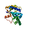

 Mass: 24.305 Da / Num. of mol.: 1 / Source method: obtained synthetically / Formula: Mg
 Mass: 18.015 Da / Num. of mol.: 376 / Source method: isolated from a natural source / Formula: H2O
 Sample preparation
 / Beamline: ID14-4 / Wavelength: 0.9756
  Processing