 Movie
Movie Controller

[English] 日本語
 Yorodumi
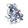
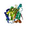
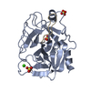
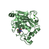
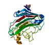
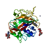
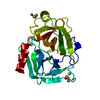
- PDB-4npr: Crystal Structure of the Family 12 Xyloglucanase from Aspergillus... -
+ Open data

- Basic information
| Entry | Database: PDB / ID: 4npr | ||||||
|---|---|---|---|---|---|---|---|
| Title | Crystal Structure of the Family 12 Xyloglucanase from Aspergillus niveus | ||||||
 Components Components | Xyloglucan-specific endo-beta-1,4-glucanase GH12 | ||||||
 Keywords Keywords | HYDROLASE / b-jelly roll / xyloglucan-specific endo-beta-1 / 4-glucanase | ||||||
| Function / homology |  Function and homology information Function and homology informationxyloglucan-specific endo-beta-1,4-glucanase / xyloglucan-specific endo-beta-1,4-glucanase activity / cellulase activity / polysaccharide catabolic process Similarity search - Function | ||||||
| Biological species |  | ||||||
| Method |  X-RAY DIFFRACTION / SYNCHROTRON / MOLECULAR REPLACEMENT / Resolution: 2.5 Å X-RAY DIFFRACTION / SYNCHROTRON / MOLECULAR REPLACEMENT / Resolution: 2.5 Å | ||||||
 Authors Authors | Cordeiro, R.L. / Santos, C.R. / Furtado, G.P. / Damasio, A.R.L. / Polizeli, M.L.T.M. / Ward, R.J. / Murakami, M.T. | ||||||
 Citation Citation | Journal: To be Published Title: Crystal Structure of the Family 12 Xyloglucanase from Aspergillus niveus Authors: Santos, C.R. / Cordeiro, R.L. / Furtado, G.P. / Damasio, A.R.L. / Polizeli, M.L.T.M. / Ward, R.J. / Murakami, M.T. | ||||||
| History |
|
- Structure visualization
| Structure viewer | Molecule: MolmilJmol/JSmol |
|---|
- Downloads & links
-Download
| PDBx/mmCIF format | 4npr.cif.gz | 94.2 KB | Display | PDBx/mmCIF format |
|---|---|---|---|---|
| PDB format | pdb4npr.ent.gz | 72.2 KB | Display | PDB format |
| PDBx/mmJSON format | 4npr.json.gz | Tree view | PDBx/mmJSON format | |
| Others |  Other downloads Other downloads |
-Validation report
| Arichive directory | https://data.pdbj.org/pub/pdb/validation_reports/np/4nprftp://data.pdbj.org/pub/pdb/validation_reports/np/4npr | HTTPS FTP |
|---|
-Related structure data
| Related structure data | 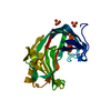 3vl8S S: Starting model for refinement |
|---|---|
| Similar structure data |


-Links
 PDBj
PDBj
- Assembly
| Deposited unit | 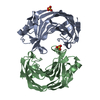
| ||||||||
|---|---|---|---|---|---|---|---|---|---|
| 1 | 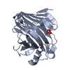
| ||||||||
| 2 | 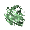
| ||||||||
| 3 | 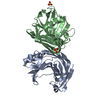
| ||||||||
| Unit cell |
|
-Components
| #1: Protein | Mass: 26039.531 Da / Num. of mol.: 2 Source method: isolated from a genetically manipulated source Source: (gene. exp.)  References: UniProt: H6TQN1, xyloglucan-specific endo-beta-1,4-glucanase #2: Chemical |   Mass: 96.063 Da / Num. of mol.: 2 / Source method: obtained synthetically / Formula: SO4 Mass: 96.063 Da / Num. of mol.: 2 / Source method: obtained synthetically / Formula: SO4#3: Water | ChemComp-HOH / |  Mass: 18.015 Da / Num. of mol.: 47 / Source method: isolated from a natural source / Formula: H2O Mass: 18.015 Da / Num. of mol.: 47 / Source method: isolated from a natural source / Formula: H2OHas protein modification | Y | |
|---|
-Experimental details
-Experiment
| Experiment | Method: X-RAY DIFFRACTION / Number of used crystals: 1 |
|---|
- Sample preparation
| Crystal | Density Matthews: 2.11 Å3/Da / Density % sol: 41.71 % |
|---|---|
| Crystal grow | Temperature: 291 K / Method: vapor diffusion, hanging drop / pH: 3.6 Details: 20% PEG 4000, 0.1M ammonium sulfate, 0.1M sodium acetate, pH 3.6, VAPOR DIFFUSION, HANGING DROP, temperature 291K |
-Data collection
| Diffraction | Mean temperature: 100 K |
|---|---|
| Diffraction source | Source: SYNCHROTRON / Site: LNLS  / Beamline: W01B-MX2 / Wavelength: 1.4586 Å / Beamline: W01B-MX2 / Wavelength: 1.4586 Å |
| Detector | Type: MARMOSAIC 225 mm CCD / Detector: CCD / Date: Jan 1, 2001 |
| Radiation | Monochromator: double-crystal Si(111) / Protocol: SINGLE WAVELENGTH / Monochromatic (M) / Laue (L): M / Scattering type: x-ray |
| Radiation wavelength | Wavelength: 1.4586 Å / Relative weight: 1 |
| Reflection | Resolution: 2.5→50 Å / Num. obs: 15884 / % possible obs: 96.9 % / Observed criterion σ(F): 2 / Observed criterion σ(I): 2 / Redundancy: 4 % / Rmerge(I) obs: 0.108 / Net I/σ(I): 11.9 |
| Reflection shell | Resolution: 2.5→2.59 Å / Redundancy: 3.5 % / Rmerge(I) obs: 0.382 / Mean I/σ(I) obs: 3.1 / Num. unique all: 1450 / % possible all: 91.9 |
- Processing
| Software |
| |||||||||||||||||||||||||||||||||||||||||||||||||||||||||||||||||||||||||||||||||||||||||||||||||||||||||
|---|---|---|---|---|---|---|---|---|---|---|---|---|---|---|---|---|---|---|---|---|---|---|---|---|---|---|---|---|---|---|---|---|---|---|---|---|---|---|---|---|---|---|---|---|---|---|---|---|---|---|---|---|---|---|---|---|---|---|---|---|---|---|---|---|---|---|---|---|---|---|---|---|---|---|---|---|---|---|---|---|---|---|---|---|---|---|---|---|---|---|---|---|---|---|---|---|---|---|---|---|---|---|---|---|---|---|
| Refinement | Method to determine structure: MOLECULAR REPLACEMENT Starting model: 3VL8 Resolution: 2.5→48.09 Å / Cor.coef. Fo:Fc: 0.94 / Cor.coef. Fo:Fc free: 0.896 / SU B: 7.821 / SU ML: 0.174 / Cross valid method: THROUGHOUT / σ(F): 2 / ESU R: 0.714 / ESU R Free: 0.295 / Stereochemistry target values: MAXIMUM LIKELIHOOD
| |||||||||||||||||||||||||||||||||||||||||||||||||||||||||||||||||||||||||||||||||||||||||||||||||||||||||
| Solvent computation | Ion probe radii: 0.8 Å / Shrinkage radii: 0.8 Å / VDW probe radii: 1.2 Å / Solvent model: MASK | |||||||||||||||||||||||||||||||||||||||||||||||||||||||||||||||||||||||||||||||||||||||||||||||||||||||||
| Displacement parameters | Biso mean: 17.941 Å2
| |||||||||||||||||||||||||||||||||||||||||||||||||||||||||||||||||||||||||||||||||||||||||||||||||||||||||
| Refinement step | Cycle: LAST / Resolution: 2.5→48.09 Å
| |||||||||||||||||||||||||||||||||||||||||||||||||||||||||||||||||||||||||||||||||||||||||||||||||||||||||
| Refine LS restraints |
| |||||||||||||||||||||||||||||||||||||||||||||||||||||||||||||||||||||||||||||||||||||||||||||||||||||||||
| LS refinement shell | Resolution: 2.499→2.564 Å / Total num. of bins used: 20
|
