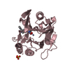
 Movie
Movie Controller


+ Open data

- Basic information
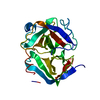
| Entry | Database: PDB / ID: 1oss | ||||||
|---|---|---|---|---|---|---|---|
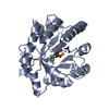
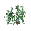
| Title | T190P STREPTOMYCES GRISEUS TRYPSIN IN COMPLEX WITH BENZAMIDINE | ||||||
 Components Components | trypsin | ||||||
 Keywords Keywords | HYDROLASE / TRYPSIN / SERINE PROTEASE / MUTANT | ||||||
| Function / homology |  Function and homology information Function and homology information | ||||||
| Biological species |  Streptomyces griseus (bacteria) Streptomyces griseus (bacteria) | ||||||
| Method |  X-RAY DIFFRACTION / RIGID BODY REFINEMENT / Resolution: 1.93 Å X-RAY DIFFRACTION / RIGID BODY REFINEMENT / Resolution: 1.93 Å | ||||||
 Authors Authors | Page, M.J. / Wong, S.L. / Hewitt, J. / Strynadka, N.C. / MacGillivray, R.T. | ||||||
 Citation Citation | Journal: Biochemistry / Year: 2003 Title: Engineering the Primary Substrate Specificity of Streptomyces griseus Trypsin. Authors: Page, M.J. / Wong, S.L. / Hewitt, J. / Strynadka, N.C. / MacGillivray, R.T. #1: Journal: J.Mol.Biol. / Year: 1988Title: Refined Crystal Structure of Streptomyces Griseus Trypsin at 1.7 Angstroms Resolution Authors: Read, R.J. / James, M.N.G. #2: Journal: Biochemistry / Year: 1984Title: Critical Comparison of Comparative Model Building of Streptomyces Griseus Trypsin Authors: Read, R.J. / Brayer, G.D. / Jurasek, L. / James, M.N.G. | ||||||
| History |
| ||||||
| Remark 999 | SEQUENCE AMINO ACID NUMBERING IS BASED ON CHYMOTRYPSINOGEN AND IS THE SAME AS THAT USED IN THE ...SEQUENCE AMINO ACID NUMBERING IS BASED ON CHYMOTRYPSINOGEN AND IS THE SAME AS THAT USED IN THE NATIVE STRUCTURE (PDB ID 1SGT). |
- Structure visualization
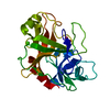
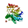
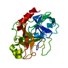
| Structure viewer | Molecule: MolmilJmol/JSmol |
|---|
- Downloads & links
-Download
| PDBx/mmCIF format | 1oss.cif.gz | 60.7 KB | Display | PDBx/mmCIF format |
|---|---|---|---|---|
| PDB format | pdb1oss.ent.gz | 43 KB | Display | PDB format |
| PDBx/mmJSON format | 1oss.json.gz | Tree view | PDBx/mmJSON format | |
| Others |  Other downloads Other downloads |
-Validation report
| Arichive directory | https://data.pdbj.org/pub/pdb/validation_reports/os/1ossftp://data.pdbj.org/pub/pdb/validation_reports/os/1oss | HTTPS FTP |
|---|
-Related structure data
| Related structure data |  1os8C  1sgtS S: Starting model for refinement C: citing same article ( |
|---|---|
| Similar structure data |


-Links
 PDBj
PDBj


- Assembly
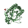
| Deposited unit | 
| ||||||||
|---|---|---|---|---|---|---|---|---|---|
| 1 |
| ||||||||
| Unit cell |
|
-Components
| #1: Protein | Mass: 23118.799 Da / Num. of mol.: 1 / Mutation: T190P Source method: isolated from a genetically manipulated source Source: (gene. exp.) Streptomyces griseus (bacteria) / Gene: SPRT / Plasmid: PWB980 / Production host: | ||||||||
|---|---|---|---|---|---|---|---|---|---|
| #2: Chemical |   Mass: 96.063 Da / Num. of mol.: 2 / Source method: obtained synthetically / Formula: SO4 Mass: 96.063 Da / Num. of mol.: 2 / Source method: obtained synthetically / Formula: SO4#3: Chemical | ChemComp-CA / |   Mass: 40.078 Da / Num. of mol.: 1 / Source method: obtained synthetically / Formula: Ca Mass: 40.078 Da / Num. of mol.: 1 / Source method: obtained synthetically / Formula: Ca#4: Chemical | ChemComp-BEN / |   Mass: 120.152 Da / Num. of mol.: 1 / Source method: obtained synthetically / Formula: C7H8N2 Mass: 120.152 Da / Num. of mol.: 1 / Source method: obtained synthetically / Formula: C7H8N2#5: Water | ChemComp-HOH / |  Mass: 18.015 Da / Num. of mol.: 193 / Source method: isolated from a natural source / Formula: H2O Mass: 18.015 Da / Num. of mol.: 193 / Source method: isolated from a natural source / Formula: H2OHas protein modification | Y | |
-Experimental details
-Experiment
| Experiment | Method: X-RAY DIFFRACTION / Number of used crystals: 1 |
|---|
- Sample preparation
| Crystal | Density Matthews: 2.02 Å3/Da / Density % sol: 38.7 % | ||||||||||||||||||||||||||||||
|---|---|---|---|---|---|---|---|---|---|---|---|---|---|---|---|---|---|---|---|---|---|---|---|---|---|---|---|---|---|---|---|
| Crystal grow | Temperature: 293 K / Method: vapor diffusion, hanging drop / pH: 6.5 Details: 1.55 M AMMONIUM SULPHATE, 10 MM CALCIUM ACETATE , pH 6.5, VAPOR DIFFUSION, HANGING DROP, temperature 293K | ||||||||||||||||||||||||||||||
| Crystal grow | *PLUS pH: 6.2 / Method: vapor diffusion, hanging drop | ||||||||||||||||||||||||||||||
| Components of the solutions | *PLUS
|
-Data collection
| Diffraction | Mean temperature: 100 K |
|---|---|
| Diffraction source | Source: ROTATING ANODE / Type: RIGAKU RU200 / Wavelength: 1.5418 Å |
| Detector | Type: MARRESEARCH / Detector: IMAGE PLATE / Date: Nov 21, 2002 |
| Radiation | Monochromator: OSMIC MIRRORS / Protocol: SINGLE WAVELENGTH / Monochromatic (M) / Laue (L): M / Scattering type: x-ray |
| Radiation wavelength | Wavelength: 1.5418 Å / Relative weight: 1 |
| Reflection | Resolution: 1.93→20 Å / Num. obs: 16130 / % possible obs: 89.7 % / Observed criterion σ(I): 2 / Biso Wilson estimate: 7.1 Å2 / Rmerge(I) obs: 0.031 |
| Reflection shell | Resolution: 1.93→2 Å / Rmerge(I) obs: 0.056 / % possible all: 79.1 |
| Reflection | *PLUS Highest resolution: 1.9 Å / Num. obs: 14471 |
| Reflection shell | *PLUS Lowest resolution: 2.05 Å / % possible obs: 79.1 % / Mean I/σ(I) obs: 25.2 |
- Processing
| Software |
| ||||||||||||||||||||||||||||||||||||
|---|---|---|---|---|---|---|---|---|---|---|---|---|---|---|---|---|---|---|---|---|---|---|---|---|---|---|---|---|---|---|---|---|---|---|---|---|---|
| Refinement | Method to determine structure: RIGID BODY REFINEMENT Starting model: 1SGT Resolution: 1.93→19.86 Å / Rfactor Rfree error: 0.008 / Isotropic thermal model: RESTRAINED / Cross valid method: THROUGHOUT / σ(F): 0 / Stereochemistry target values: Engh & Huber Details: RESIDUES 165 AND 192 HAVE TWO ALTERNATE CONFORMATIONS THAT ARE CLEARLY VISIBLE IN THE ELECTRON DENSITY
| ||||||||||||||||||||||||||||||||||||
| Solvent computation | Solvent model: FLAT MODEL / Bsol: 56.6589 Å2 / ksol: 0.442522 e/Å3 | ||||||||||||||||||||||||||||||||||||
| Displacement parameters | Biso mean: 12.8 Å2
| ||||||||||||||||||||||||||||||||||||
| Refine analyze | Luzzati coordinate error free: 0.23 Å / Luzzati sigma a free: 0.12 Å | ||||||||||||||||||||||||||||||||||||
| Refinement step | Cycle: LAST / Resolution: 1.93→19.86 Å
| ||||||||||||||||||||||||||||||||||||
| Refine LS restraints |
| ||||||||||||||||||||||||||||||||||||
| LS refinement shell | Resolution: 1.93→2.05 Å / Rfactor Rfree error: 0.021 / Total num. of bins used: 6
| ||||||||||||||||||||||||||||||||||||
| Xplor file |
| ||||||||||||||||||||||||||||||||||||
| Software | *PLUS Name: XTALVIEW / Classification: refinement | ||||||||||||||||||||||||||||||||||||
| Refinement | *PLUS Highest resolution: 1.9 Å | ||||||||||||||||||||||||||||||||||||
| Solvent computation | *PLUS | ||||||||||||||||||||||||||||||||||||
| Displacement parameters | *PLUS | ||||||||||||||||||||||||||||||||||||
| Refine LS restraints | *PLUS
| ||||||||||||||||||||||||||||||||||||
| LS refinement shell | *PLUS Rfactor Rfree: 0.2 |
