 Movie
Movie Controller

[English] 日本語
 Yorodumi
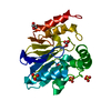
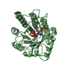
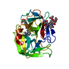
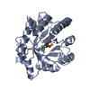
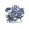
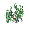
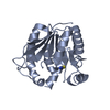
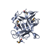
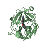
- PDB-1x8g: Crystal Structure of the Mono-Zinc Carbapenemase CphA from Aeromo... -
+ Open data

- Basic information
| Entry | Database: PDB / ID: 1x8g | ||||||
|---|---|---|---|---|---|---|---|
| Title | Crystal Structure of the Mono-Zinc Carbapenemase CphA from Aeromonas Hydrophyla | ||||||
 Components Components | Beta-lactamase | ||||||
 Keywords Keywords | HYDROLASE | ||||||
| Function / homology |  Function and homology information Function and homology informationantibiotic catabolic process / beta-lactamase activity / beta-lactamase / periplasmic space / response to antibiotic / zinc ion binding Similarity search - Function | ||||||
| Biological species |  Aeromonas hydrophila (bacteria) Aeromonas hydrophila (bacteria) | ||||||
| Method |  X-RAY DIFFRACTION / SYNCHROTRON / MOLECULAR REPLACEMENT / Resolution: 1.7 Å X-RAY DIFFRACTION / SYNCHROTRON / MOLECULAR REPLACEMENT / Resolution: 1.7 Å | ||||||
 Authors Authors | Garau, G. / Dideberg, O. | ||||||
 Citation Citation | Journal: J.Mol.Biol. / Year: 2005 Title: A Metallo-beta-lactamase Enzyme in Action: Crystal Structures of the Monozinc Carbapenemase CphA and its Complex with Biapenem Authors: Garau, G. / Bebrone, C. / Anne, C. / Galleni, M. / Frere, J.M. / Dideberg, O. #1: Journal: Antimicrob.Agents Chemother. / Year: 2004 Title: Update of the standard numbering scheme for class B beta-lactamases Authors: Garau, G. / Garcia-Saez, I. / Bebrone, C. / Anne, C. / Mercuri, P. / Galleni, M. / Frere, J.M. / Dideberg, O. #2: Journal: Embo J. / Year: 1995Title: The 3-D structure of a zinc metallo-beta-lactamase from Bacillus cereus reveals a new type of protein fold Authors: Carfi, A. / Pares, S. / Duee, E. / Galleni, M. / Duez, C. / Frere, J.M. / Dideberg, O. #3: Journal: J.Mol.Biol. / Year: 2003Title: Three-dimensional structure of FEZ-1, a monomeric subclass B3 metallo-beta-lactamase from Fluoribacter gormanii, in native form and in complex with D-captopril Authors: Garcia-Saez, I. / Mercuri, P.S. / Papamicael, C. / Kahn, R. / Frere, J.M. / Galleni, M. / Rossolini, G.M. / Dideberg, O. | ||||||
| History |
|
- Structure visualization
| Structure viewer | Molecule: MolmilJmol/JSmol |
|---|
- Downloads & links
-Download
| PDBx/mmCIF format | 1x8g.cif.gz | 63.4 KB | Display | PDBx/mmCIF format |
|---|---|---|---|---|
| PDB format | pdb1x8g.ent.gz | 45.6 KB | Display | PDB format |
| PDBx/mmJSON format | 1x8g.json.gz | Tree view | PDBx/mmJSON format | |
| Others |  Other downloads Other downloads |
-Validation report
| Arichive directory | https://data.pdbj.org/pub/pdb/validation_reports/x8/1x8gftp://data.pdbj.org/pub/pdb/validation_reports/x8/1x8g | HTTPS FTP |
|---|
-Related structure data


-Links
 PDBj
PDBj


- Assembly
| Deposited unit | 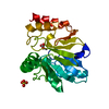
| ||||||||
|---|---|---|---|---|---|---|---|---|---|
| 1 |
| ||||||||
| Unit cell |
|
-Components
| #1: Protein | Mass: 25220.818 Da / Num. of mol.: 1 Source method: isolated from a genetically manipulated source Source: (gene. exp.) Aeromonas hydrophila (bacteria) / Gene: cphA / Plasmid: PUC20-CPHA / Production host: |
|---|---|
| #2: Chemical | ChemComp-ZN /   Mass: 65.409 Da / Num. of mol.: 1 / Source method: obtained synthetically / Formula: Zn Mass: 65.409 Da / Num. of mol.: 1 / Source method: obtained synthetically / Formula: Zn |
| #3: Chemical | ChemComp-CO3 /   Mass: 60.009 Da / Num. of mol.: 1 / Source method: obtained synthetically / Formula: CO3 Mass: 60.009 Da / Num. of mol.: 1 / Source method: obtained synthetically / Formula: CO3 |
| #4: Chemical | ChemComp-SO4 /   Mass: 96.063 Da / Num. of mol.: 1 / Source method: obtained synthetically / Formula: SO4 Mass: 96.063 Da / Num. of mol.: 1 / Source method: obtained synthetically / Formula: SO4 |
| #5: Water | ChemComp-HOH /  Mass: 18.015 Da / Num. of mol.: 178 / Source method: isolated from a natural source / Formula: H2O Mass: 18.015 Da / Num. of mol.: 178 / Source method: isolated from a natural source / Formula: H2O |
| Has protein modification | N |
-Experimental details
-Experiment
| Experiment | Method: X-RAY DIFFRACTION / Number of used crystals: 1 |
|---|
- Sample preparation
| Crystal | Density Matthews: 2.5 Å3/Da / Density % sol: 51 % |
|---|---|
| Crystal grow | Temperature: 278 K / Method: vapor diffusion, hanging drop / pH: 6.5 Details: PEG, AS, pH 6.50, VAPOR DIFFUSION, HANGING DROP, temperature 278K |
-Data collection
| Diffraction | Mean temperature: 100 K |
|---|---|
| Diffraction source | Source: SYNCHROTRON / Site: ESRF  / Beamline: BM30A / Wavelength: 0.979469 / Wavelength: 0.979469 Å / Beamline: BM30A / Wavelength: 0.979469 / Wavelength: 0.979469 Å |
| Detector | Type: MARRESEARCH / Detector: CCD / Date: Jun 18, 2003 |
| Radiation | Monochromator: SI 111 / Protocol: SINGLE WAVELENGTH / Monochromatic (M) / Laue (L): M / Scattering type: x-ray |
| Radiation wavelength | Wavelength: 0.979469 Å / Relative weight: 1 |
| Reflection | Resolution: 1.7→58.722 Å / Num. obs: 27567 / % possible obs: 94.7 % / Redundancy: 3.5 % / Rmerge(I) obs: 0.123 / Rsym value: 0.123 / Net I/σ(I): 3.9 |
| Reflection shell | Resolution: 1.7→1.79 Å / Redundancy: 3 % / Rmerge(I) obs: 0.312 / Mean I/σ(I) obs: 2.8 / Rsym value: 0.312 / % possible all: 94.7 |
- Processing
| Software |
| |||||||||||||||||||||||||||||||||||||||||||||||||||||||||||||||||||||||||||||||||||||||||||||||||||||||||||||||||||
|---|---|---|---|---|---|---|---|---|---|---|---|---|---|---|---|---|---|---|---|---|---|---|---|---|---|---|---|---|---|---|---|---|---|---|---|---|---|---|---|---|---|---|---|---|---|---|---|---|---|---|---|---|---|---|---|---|---|---|---|---|---|---|---|---|---|---|---|---|---|---|---|---|---|---|---|---|---|---|---|---|---|---|---|---|---|---|---|---|---|---|---|---|---|---|---|---|---|---|---|---|---|---|---|---|---|---|---|---|---|---|---|---|---|---|---|---|
| Refinement | Method to determine structure: MOLECULAR REPLACEMENT Starting model: CphA (N220G mutant) Resolution: 1.7→25 Å / Cor.coef. Fo:Fc: 0.939 / Cor.coef. Fo:Fc free: 0.937 / SU B: 1.915 / SU ML: 0.063 / Cross valid method: THROUGHOUT / ESU R: 0.108 / ESU R Free: 0.098 / Stereochemistry target values: MAXIMUM LIKELIHOOD / Details: HYDROGENS HAVE BEEN ADDED IN THE RIDING POSITIONS
| |||||||||||||||||||||||||||||||||||||||||||||||||||||||||||||||||||||||||||||||||||||||||||||||||||||||||||||||||||
| Solvent computation | Ion probe radii: 0.8 Å / Shrinkage radii: 0.8 Å / VDW probe radii: 1.4 Å / Solvent model: BABINET MODEL WITH MASK | |||||||||||||||||||||||||||||||||||||||||||||||||||||||||||||||||||||||||||||||||||||||||||||||||||||||||||||||||||
| Displacement parameters | Biso mean: 14.394 Å2
| |||||||||||||||||||||||||||||||||||||||||||||||||||||||||||||||||||||||||||||||||||||||||||||||||||||||||||||||||||
| Refinement step | Cycle: LAST / Resolution: 1.7→25 Å
| |||||||||||||||||||||||||||||||||||||||||||||||||||||||||||||||||||||||||||||||||||||||||||||||||||||||||||||||||||
| Refine LS restraints |
| |||||||||||||||||||||||||||||||||||||||||||||||||||||||||||||||||||||||||||||||||||||||||||||||||||||||||||||||||||
| LS refinement shell | Resolution: 1.7→1.744 Å / Total num. of bins used: 20 /
|
