 Movie
Movie Controller

[English] 日本語
 Yorodumi
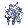
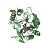
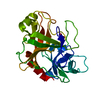
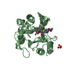
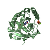
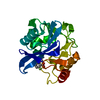
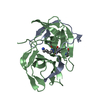
- PDB-1sgt: REFINED CRYSTAL STRUCTURE OF STREPTOMYCES GRISEUS TRYPSIN AT 1.7 ... -
+ Open data

- Basic information
| Entry | Database: PDB / ID: 1sgt | ||||||
|---|---|---|---|---|---|---|---|
| Title | REFINED CRYSTAL STRUCTURE OF STREPTOMYCES GRISEUS TRYPSIN AT 1.7 ANGSTROMS RESOLUTION | ||||||
 Components Components | TRYPSIN | ||||||
 Keywords Keywords | HYDROLASE (SERINE PROTEINASE) | ||||||
| Function / homology |  Function and homology information Function and homology information | ||||||
| Biological species |  Streptomyces griseus (bacteria) Streptomyces griseus (bacteria) | ||||||
| Method |  X-RAY DIFFRACTION / Resolution: 1.7 Å X-RAY DIFFRACTION / Resolution: 1.7 Å | ||||||
 Authors Authors | Read, R.J. / James, M.N.G. | ||||||
 Citation Citation | Journal: J.Mol.Biol. / Year: 1988 Title: Refined crystal structure of Streptomyces griseus trypsin at 1.7 A resolution. Authors: Read, R.J. / James, M.N. #1: Journal: Biochemistry / Year: 1984Title: Critical Comparison of Comparative Model Building of Streptomyces Griseus Trypsin Authors: Read, R.J. / Brayer, G.D. / Jurasek, L. / James, M.N.G. | ||||||
| History |
| ||||||
| Remark 700 | SHEET SHEETS S1 AND S2 ARE BOTH SIX-STRANDED SHEETS FORMING A BETA-BARREL. THIS IS REPRESENTED ON ...SHEET SHEETS S1 AND S2 ARE BOTH SIX-STRANDED SHEETS FORMING A BETA-BARREL. THIS IS REPRESENTED ON THE SHEET RECORDS BELOW BY A SEVEN-STRANDED SHEET WITH THE FIRST AND LAST STRANDS IDENTICAL. |
- Structure visualization
| Structure viewer | Molecule: MolmilJmol/JSmol |
|---|
- Downloads & links
-Download
| PDBx/mmCIF format | 1sgt.cif.gz | 58.6 KB | Display | PDBx/mmCIF format |
|---|---|---|---|---|
| PDB format | pdb1sgt.ent.gz | 41.5 KB | Display | PDB format |
| PDBx/mmJSON format | 1sgt.json.gz | Tree view | PDBx/mmJSON format | |
| Others |  Other downloads Other downloads |
-Validation report
| Arichive directory | https://data.pdbj.org/pub/pdb/validation_reports/sg/1sgtftp://data.pdbj.org/pub/pdb/validation_reports/sg/1sgt | HTTPS FTP |
|---|
-Related structure data
| Similar structure data |
|---|


-Links
 PDBj
PDBj

- Assembly
| Deposited unit | 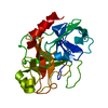
| ||||||||
|---|---|---|---|---|---|---|---|---|---|
| 1 |
| ||||||||
| Unit cell |
| ||||||||
| Atom site foot note | 1: THR 20 - POSSIBLE STATIC DISORDER WITH CHI 1 ROTATED BY -120 DEGREES. 2: ARG 21 - POOR DENSITY FOR GUANIDINIUM GROUP. 3: THR 65 - POSSIBLE STATIC DISORDER WITH CHI 1 ROTATED BY +120 DEGREES. 4: LYS 82 - GOOD DENSITY UP TO NZ WHICH IS NOT VISIBLE. / 5: ARG 84 - NOISY DENSITY BEYOND CD. / 6: LYS 87 - VERY WEAK DENSITY BEYOND CD. 7: THR 98 - POSSIBLE STATIC DISORDER WITH CHI 1 ROTATED BY +120 DEGREES. 8: GLN 110 - NOISY SIDE CHAIN DENSITY. / 9: LYS 122 - WEAK DENSITY BEYOND CG. 10: GLN 133 - CHI 3 ANGLE DIFFICULT TO ESTABLISH FROM DENSITY. 11: ARG 145 - NOISY AND AMBIGUOUS SIDE CHAIN DENSITY, PROJECTING INTO SOLVENT. 12: ASN 174 - LITTLE DENSITY FOR OD1 AND ND2. / 13: GLN 192 - DENSITY NOISY AND WEAK BEYOND CB. 14: ASN 204 - NOISY DENSITY, POSSIBLY DUE TO STATIC DISORDER AROUND CHI 1. 15: ASP 205 - VERY WEAK DENSITY FOR CB, NOISY DENSITY FOR CARBOXYL GROUP. 16: SER 236 - POSSIBLE STATIC DISORDER ABOUT CHI 1. 17: ARG 243 - VERY NOISY DENSITY, SIDE CHAIN PAST CG IS ESSENTIALLY ARBITRARY. | ||||||||
| Components on special symmetry positions |
|
-Components
| #1: Protein | Mass: 23076.762 Da / Num. of mol.: 1 Source method: isolated from a genetically manipulated source Source: (gene. exp.) Streptomyces griseus (bacteria) / References: UniProt: P00775, trypsin |
|---|---|
| #2: Chemical | ChemComp-CA /   Mass: 40.078 Da / Num. of mol.: 1 / Source method: obtained synthetically / Formula: Ca Mass: 40.078 Da / Num. of mol.: 1 / Source method: obtained synthetically / Formula: Ca |
| #3: Water | ChemComp-HOH /  Mass: 18.015 Da / Num. of mol.: 192 / Source method: isolated from a natural source / Formula: H2O Mass: 18.015 Da / Num. of mol.: 192 / Source method: isolated from a natural source / Formula: H2O |
| Has protein modification | Y |
| Source details | SGT IS PURIFIED FROM PRONASE, A COMMERCIAL PRODUCT OBTAINED FROM THE EXTRACELLULAR CULTURE FILTRATE ...SGT IS PURIFIED FROM PRONASE, A COMMERCIAL |
-Experimental details
-Experiment
| Experiment | Method: X-RAY DIFFRACTION |
|---|
- Sample preparation
| Crystal | Density Matthews: 2.4 Å3/Da / Density % sol: 48.67 % | ||||||||||||||||||||||||||||||
|---|---|---|---|---|---|---|---|---|---|---|---|---|---|---|---|---|---|---|---|---|---|---|---|---|---|---|---|---|---|---|---|
| Crystal grow | *PLUS pH: 6.2 / Method: microdialysis | ||||||||||||||||||||||||||||||
| Components of the solutions | *PLUS
|
-Data collection
| Reflection | *PLUS Highest resolution: 1.7 Å / Num. obs: 24878 / Num. measured all: 25580 |
|---|---|
| Reflection shell | *PLUS |
- Processing
| Software | Name: PROLSQ / Classification: refinement | ||||||||||||||||||||||||||||||||||||||||||||||||||||||||||||||||||||||||||||||||||||
|---|---|---|---|---|---|---|---|---|---|---|---|---|---|---|---|---|---|---|---|---|---|---|---|---|---|---|---|---|---|---|---|---|---|---|---|---|---|---|---|---|---|---|---|---|---|---|---|---|---|---|---|---|---|---|---|---|---|---|---|---|---|---|---|---|---|---|---|---|---|---|---|---|---|---|---|---|---|---|---|---|---|---|---|---|---|
| Refinement | Resolution: 1.7→8 Å / σ(I): 0
| ||||||||||||||||||||||||||||||||||||||||||||||||||||||||||||||||||||||||||||||||||||
| Refinement step | Cycle: LAST / Resolution: 1.7→8 Å
| ||||||||||||||||||||||||||||||||||||||||||||||||||||||||||||||||||||||||||||||||||||
| Refine LS restraints |
| ||||||||||||||||||||||||||||||||||||||||||||||||||||||||||||||||||||||||||||||||||||
| Refinement | *PLUS σ(I): 1 / Highest resolution: 1.7 Å / Lowest resolution: 8 Å / Num. reflection obs: 20046 / Rfactor obs: 0.161 | ||||||||||||||||||||||||||||||||||||||||||||||||||||||||||||||||||||||||||||||||||||
| Solvent computation | *PLUS | ||||||||||||||||||||||||||||||||||||||||||||||||||||||||||||||||||||||||||||||||||||
| Displacement parameters | *PLUS |
