 Movie
Movie Controller

[English] 日本語
 Yorodumi
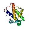
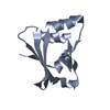
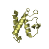
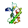
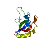
- PDB-7ax5: Anammox-specific acyl carrier protein from Kuenenia stuttgartiens... -
+ Open data

- Basic information
| Entry | Database: PDB / ID: 7ax5 | ||||||
|---|---|---|---|---|---|---|---|
| Title | Anammox-specific acyl carrier protein from Kuenenia stuttgartiensis; ensemble refinement | ||||||
 Components Components | Similar to acyl carrier protein | ||||||
 Keywords Keywords | LIPID BINDING PROTEIN / anammox / ladderane / acyl carrier protein / ensemble refinement | ||||||
| Function / homology | Phosphopantetheine attachment site / ACP-like superfamily / Carrier protein (CP) domain profile. / Phosphopantetheine binding ACP domain / metal ion binding / Putative acyl carrier protein Function and homology information Function and homology information | ||||||
| Biological species |  Kuenenia stuttgartiensis (bacteria) Kuenenia stuttgartiensis (bacteria) | ||||||
| Method |  X-RAY DIFFRACTION / SYNCHROTRON / SAD / Resolution: 1.756 Å X-RAY DIFFRACTION / SYNCHROTRON / SAD / Resolution: 1.756 Å | ||||||
 Authors Authors | Dietl, A. / Barends, T. | ||||||
| Funding support | European Union, 1items
| ||||||
 Citation Citation | Journal: Proteins / Year: 2022 Title: Dynamics in an unusual acyl carrier protein from a ladderane lipid-synthesizing organism. Authors: Dietl, A. / Barends, T.R.M. | ||||||
| History |
|
- Structure visualization
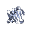
| Structure viewer | Molecule: MolmilJmol/JSmol |
|---|
- Downloads & links
-Download
| PDBx/mmCIF format | 7ax5.cif.gz | 4.9 MB | Display | PDBx/mmCIF format |
|---|---|---|---|---|
| PDB format | pdb7ax5.ent.gz | 4.2 MB | Display | PDB format |
| PDBx/mmJSON format | 7ax5.json.gz | Tree view | PDBx/mmJSON format | |
| Others |  Other downloads Other downloads |
-Validation report
| Summary document | 7ax5_validation.pdf.gz | 1 MB | Display | wwPDB validaton report |
|---|---|---|---|---|
| Full document | 7ax5_full_validation.pdf.gz | 1.2 MB | Display | |
| Data in XML | 7ax5_validation.xml.gz | 99.7 KB | Display | |
| Data in CIF | 7ax5_validation.cif.gz | 244.2 KB | Display | |
| Arichive directory | https://data.pdbj.org/pub/pdb/validation_reports/ax/7ax5ftp://data.pdbj.org/pub/pdb/validation_reports/ax/7ax5 | HTTPS FTP |
-Related structure data


-Links
 PDBj
PDBj
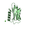
- Assembly
| Deposited unit | 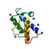
| ||||||||
|---|---|---|---|---|---|---|---|---|---|
| 1 |
| ||||||||
| Unit cell |
| ||||||||
| Number of models | 75 |
-Components
| #1: Protein | Mass: 10732.137 Da / Num. of mol.: 1 Source method: isolated from a genetically manipulated source Source: (gene. exp.) Kuenenia stuttgartiensis (bacteria) / Gene: acpP, acpP_2, KSMBR1_3472, kuste3603 / Production host: | ||||
|---|---|---|---|---|---|
| #2: Chemical | ChemComp-ZN /   Mass: 65.409 Da / Num. of mol.: 7 / Source method: obtained synthetically / Formula: Zn Mass: 65.409 Da / Num. of mol.: 7 / Source method: obtained synthetically / Formula: Zn#3: Water | ChemComp-HOH / |  Mass: 18.015 Da / Num. of mol.: 16 / Source method: isolated from a natural source / Formula: H2O Mass: 18.015 Da / Num. of mol.: 16 / Source method: isolated from a natural source / Formula: H2OHas ligand of interest | N | |
-Experimental details
-Experiment
| Experiment | Method: X-RAY DIFFRACTION / Number of used crystals: 1 |
|---|
- Sample preparation
| Crystal | Density Matthews: 2.45 Å3/Da / Density % sol: 49.83 % |
|---|---|
| Crystal grow | Temperature: 293 K / Method: vapor diffusion, hanging drop Details: 28% (v/v) PEG 400, 200 mM calcium acetate, 100 mM sodium acetate pH 4.5 and 10 mM zinc chloride |
-Data collection
| Diffraction | Mean temperature: 100 K / Serial crystal experiment: N |
|---|---|
| Diffraction source | Source: SYNCHROTRON / Site: SLS  / Beamline: X10SA / Wavelength: 1 Å / Beamline: X10SA / Wavelength: 1 Å |
| Detector | Type: DECTRIS PILATUS3 S 6M / Detector: PIXEL / Date: Aug 28, 2015 |
| Radiation | Protocol: SINGLE WAVELENGTH / Monochromatic (M) / Laue (L): M / Scattering type: x-ray |
| Radiation wavelength | Wavelength: 1 Å / Relative weight: 1 |
| Reflection | Resolution: 1.756→38.404 Å / Num. obs: 7492 / % possible obs: 91.6 % / Redundancy: 5.3 % / Rpim(I) all: 0.025 / Net I/σ(I): 16.4 |
| Reflection shell | Resolution: 1.76→1.9 Å / Num. unique obs: 1132 / Rpim(I) all: 0.652 |
- Processing
| Software |
| ||||||||||||||||||||||||
|---|---|---|---|---|---|---|---|---|---|---|---|---|---|---|---|---|---|---|---|---|---|---|---|---|---|
| Refinement | Method to determine structure: SAD / Resolution: 1.756→38.404 Å / SU ML: 0.16 / Cross valid method: THROUGHOUT / σ(F): 1.34 / Phase error: 38.32 / Stereochemistry target values: ML
| ||||||||||||||||||||||||
| Solvent computation | Shrinkage radii: 0.8 Å / VDW probe radii: 1 Å / Solvent model: FLAT BULK SOLVENT MODEL | ||||||||||||||||||||||||
| Displacement parameters | Biso min: 99999 Å2 | ||||||||||||||||||||||||
| Refinement step | Cycle: final / Resolution: 1.756→38.404 Å
| ||||||||||||||||||||||||
| LS refinement shell | Refine-ID: X-RAY DIFFRACTION / Rfactor Rfree error: 0
|
