 Movie
Movie Controller

[English] 日本語
 Yorodumi
- PDB-7ask: 43S preinitiation complex from Trypanosoma cruzi with the kDDX60 ... -
+ Open data

- Basic information
| Entry | Database: PDB / ID: 7ask | |||||||||||||||
|---|---|---|---|---|---|---|---|---|---|---|---|---|---|---|---|---|
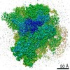
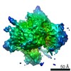
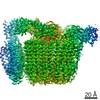
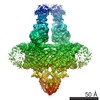
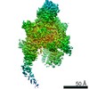
| Title | 43S preinitiation complex from Trypanosoma cruzi with the kDDX60 helicase bound with ATP | |||||||||||||||
 Components Components | kDDX60 | |||||||||||||||
 Keywords Keywords | TRANSLATION / preinitiation / factors / kinetoplastid / helicase | |||||||||||||||
| Function / homology |  Function and homology information Function and homology informationhelicase activity / nucleic acid binding / hydrolase activity / ATP binding / cytoplasm Similarity search - Function | |||||||||||||||
| Biological species |  | |||||||||||||||
| Method | ELECTRON MICROSCOPY / single particle reconstruction / cryo EM / Resolution: 4.3 Å | |||||||||||||||
 Authors Authors | Bochler, A. / Brito Querido, J. / Prilepskaja, T. / Soufari, H. / Del Cistia, M.L. / Kuhn, L. / Rimoldi Ribeiro, A. / Valasek, L.S. / Hashem, Y. | |||||||||||||||
| Funding support |  France, France,  Czech Republic, 4items Czech Republic, 4items
| |||||||||||||||
 Citation Citation | Journal: Cell Rep / Year: 2020 Title: Structural Differences in Translation Initiation between Pathogenic Trypanosomatids and Their Mammalian Hosts. Authors: Anthony Bochler / Jailson Brito Querido / Terezie Prilepskaja / Heddy Soufari / Angelita Simonetti / Mayara Lucia Del Cistia / Lauriane Kuhn / Aline Rimoldi Ribeiro / Leoš Shivaya Valášek / Yaser Hashem / Abstract: Canonical mRNA translation in eukaryotes begins with the formation of the 43S pre-initiation complex (PIC). Its assembly requires binding of initiator Met-tRNA and several eukaryotic initiation ...Canonical mRNA translation in eukaryotes begins with the formation of the 43S pre-initiation complex (PIC). Its assembly requires binding of initiator Met-tRNA and several eukaryotic initiation factors (eIFs) to the small ribosomal subunit (40S). Compared to their mammalian hosts, trypanosomatids present significant structural differences in their 40S, suggesting substantial variability in translation initiation. Here, we determine the structure of the 43S PIC from Trypanosoma cruzi, the parasite causing Chagas disease. Our structure shows numerous specific features, such as the variant eIF3 structure and its unique interactions with the large rRNA expansion segments (ESs) 9, 7, and 6, and the association of a kinetoplastid-specific DDX60-like helicase. It also reveals the 40S-binding site of the eIF5 C-terminal domain and structures of key terminal tails of several conserved eIFs underlying their activities within the PIC. Our results are corroborated by glutathione S-transferase (GST) pull-down assays in both human and T. cruzi and mass spectrometry data. | |||||||||||||||
| History |
|
- Structure visualization
| Movie |
Movie viewer |
|---|---|
| Structure viewer | Molecule: MolmilJmol/JSmol |
- Downloads & links
-Download
| PDBx/mmCIF format | 7ask.cif.gz | 299 KB | Display | PDBx/mmCIF format |
|---|---|---|---|---|
| PDB format | pdb7ask.ent.gz | 217 KB | Display | PDB format |
| PDBx/mmJSON format | 7ask.json.gz | Tree view | PDBx/mmJSON format | |
| Others |  Other downloads Other downloads |
-Validation report
| Arichive directory | https://data.pdbj.org/pub/pdb/validation_reports/as/7askftp://data.pdbj.org/pub/pdb/validation_reports/as/7ask | HTTPS FTP |
|---|
-Related structure data
| Related structure data |  11895MC  7aseC C: citing same article ( M: map data used to model this data |
|---|---|
| Similar structure data |


-Links
 PDBj
PDBj
- Assembly
| Deposited unit | 
|
|---|---|
| 1 |
|
-Components
| #1: Protein | Mass: 246749.875 Da / Num. of mol.: 1 / Source method: isolated from a natural source / Source: (natural) |
|---|---|
| #2: Chemical | ChemComp-MG /   Mass: 24.305 Da / Num. of mol.: 1 / Source method: obtained synthetically / Formula: Mg Mass: 24.305 Da / Num. of mol.: 1 / Source method: obtained synthetically / Formula: Mg |
| #3: Chemical | ChemComp-ADP /   Mass: 427.201 Da / Num. of mol.: 1 / Source method: obtained synthetically / Formula: C10H15N5O10P2 / Comment: ADP, energy-carrying molecule*YM Mass: 427.201 Da / Num. of mol.: 1 / Source method: obtained synthetically / Formula: C10H15N5O10P2 / Comment: ADP, energy-carrying molecule*YM |
| Has ligand of interest | N |
-Experimental details
-Experiment
| Experiment | Method: ELECTRON MICROSCOPY |
|---|---|
| EM experiment | Aggregation state: PARTICLE / 3D reconstruction method: single particle reconstruction |
- Sample preparation
| Component | Name: 43S preinitiation complex from Trypanosoma cruzi with the helicase kDDX60 Type: COMPLEX / Entity ID: #1 / Source: NATURAL |
|---|---|
| Source (natural) | Organism: |
| Buffer solution | pH: 7.4 |
| Specimen | Embedding applied: NO / Shadowing applied: NO / Staining applied: NO / Vitrification applied: YES |
| Vitrification | Instrument: FEI VITROBOT MARK IV / Cryogen name: ETHANE / Humidity: 100 % / Chamber temperature: 277.15 K |
- Electron microscopy imaging
| Experimental equipment |  Model: Titan Krios / Image courtesy: FEI Company |
|---|---|
| Microscopy | Model: FEI TITAN KRIOS |
| Electron gun | Electron source:  FIELD EMISSION GUN / Accelerating voltage: 300 kV / Illumination mode: FLOOD BEAM FIELD EMISSION GUN / Accelerating voltage: 300 kV / Illumination mode: FLOOD BEAM |
| Electron lens | Mode: OTHER |
| Image recording | Electron dose: 30 e/Å2 / Film or detector model: GATAN K2 SUMMIT (4k x 4k) |
- Processing
| CTF correction | Type: NONE |
|---|---|
| 3D reconstruction | Resolution: 4.3 Å / Resolution method: FSC 0.143 CUT-OFF / Num. of particles: 19700 / Symmetry type: POINT |
