 Movie
Movie Controller


+ Open data

- Basic information
| Entry | Database: PDB / ID: 6jiv | ||||||
|---|---|---|---|---|---|---|---|
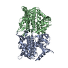
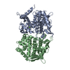
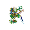
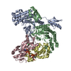
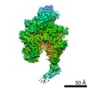
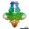
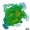
| Title | SspE crystal structure | ||||||
 Components Components | SspE protein | ||||||
 Keywords Keywords | HYDROLASE / DNase / PT | ||||||
| Biological species |  Streptomyces yokosukanensis (bacteria) Streptomyces yokosukanensis (bacteria) | ||||||
| Method |  X-RAY DIFFRACTION / SYNCHROTRON / SAD / Resolution: 3.31 Å X-RAY DIFFRACTION / SYNCHROTRON / SAD / Resolution: 3.31 Å | ||||||
 Authors Authors | Bing, Y.Z. / Yang, H.G. | ||||||
 Citation Citation | Journal: Nat Microbiol / Year: 2020 Title: SspABCD-SspE is a phosphorothioation-sensing bacterial defence system with broad anti-phage activities. Authors: Xiong, X. / Wu, G. / Wei, Y. / Liu, L. / Zhang, Y. / Su, R. / Jiang, X. / Li, M. / Gao, H. / Tian, X. / Zhang, Y. / Hu, L. / Chen, S. / Tang, Y. / Jiang, S. / Huang, R. / Li, Z. / Wang, Y. / ...Authors: Xiong, X. / Wu, G. / Wei, Y. / Liu, L. / Zhang, Y. / Su, R. / Jiang, X. / Li, M. / Gao, H. / Tian, X. / Zhang, Y. / Hu, L. / Chen, S. / Tang, Y. / Jiang, S. / Huang, R. / Li, Z. / Wang, Y. / Deng, Z. / Wang, J. / Dedon, P.C. / Chen, S. / Wang, L. | ||||||
| History |
|
- Structure visualization
| Structure viewer | Molecule:  MolmilJmol/JSmol MolmilJmol/JSmol |
|---|
- Downloads & links
-Download
| PDBx/mmCIF format | 6jiv.cif.gz | 415.4 KB | Display | PDBx/mmCIF format |
|---|---|---|---|---|
| PDB format | pdb6jiv.ent.gz | 317.5 KB | Display | PDB format |
| PDBx/mmJSON format | 6jiv.json.gz | Tree view | PDBx/mmJSON format | |
| Others |  Other downloads Other downloads |
-Validation report
| Arichive directory | https://data.pdbj.org/pub/pdb/validation_reports/ji/6jivftp://data.pdbj.org/pub/pdb/validation_reports/ji/6jiv | HTTPS FTP |
|---|
-Related structure data

-Links
 PDBj
PDBj- Assembly
| Deposited unit | 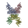
| ||||||||
|---|---|---|---|---|---|---|---|---|---|
| 1 |
| ||||||||
| Unit cell |
|
-Components
| #1: Protein | Mass: 87803.883 Da / Num. of mol.: 4 / Source method: isolated from a natural source / Source: (natural) Streptomyces yokosukanensis (bacteria)Has ligand of interest | N | Has protein modification | Y | |
|---|
-Experimental details
-Experiment
| Experiment | Method: X-RAY DIFFRACTION / Number of used crystals: 1 |
|---|
- Sample preparation
| Crystal | Density Matthews: 3.21 Å3/Da / Density % sol: 61.66 % Description: The entry contains friedel pairs in F_plus/minus columns and I_plus/minus columns. |
|---|---|
| Crystal grow | Temperature: 287.15 K / Method: vapor diffusion, hanging drop / pH: 7 / Details: PEG MME 5000, HEPES pH 7.0, Tacsimate pH 7.0 |
-Data collection
| Diffraction | Mean temperature: 298 K / Serial crystal experiment: N |
|---|---|
| Diffraction source | Source: SYNCHROTRON / Site: SSRF  / Beamline: BL19U1 / Wavelength: 0.9789 Å / Beamline: BL19U1 / Wavelength: 0.9789 Å |
| Detector | Type: DECTRIS PILATUS3 2M / Detector: PIXEL / Date: Jun 30, 2018 |
| Radiation | Protocol: MAD / Monochromatic (M) / Laue (L): M / Scattering type: x-ray |
| Radiation wavelength | Wavelength: 0.9789 Å / Relative weight: 1 |
| Reflection | Resolution: 3.31→125.33 Å / Num. obs: 64318 / % possible obs: 99.5 % / Redundancy: 6.9 % / CC1/2: 0.998 / Net I/σ(I): 8.58 |
| Reflection shell | Resolution: 3.3→3.5 Å / Num. unique obs: 52096 / CC1/2: 0.998 |
- Processing
| Software |
| ||||||||||||||||||||||||||||||||||||||||||||||||||||||||||||
|---|---|---|---|---|---|---|---|---|---|---|---|---|---|---|---|---|---|---|---|---|---|---|---|---|---|---|---|---|---|---|---|---|---|---|---|---|---|---|---|---|---|---|---|---|---|---|---|---|---|---|---|---|---|---|---|---|---|---|---|---|---|
| Refinement | Method to determine structure: SAD / Resolution: 3.31→125.33 Å / Cor.coef. Fo:Fc: 0.802 / Cor.coef. Fo:Fc free: 0.76 / Cross valid method: THROUGHOUT / σ(F): 0 / ESU R: 2.678 / ESU R Free: 0.711 / Stereochemistry target values: MAXIMUM LIKELIHOOD Details: The entry contains friedel pairs in F_plus/minus columns and I_plus/minus columns. HYDROGENS HAVE BEEN ADDED IN THE RIDING POSITIONS U VALUES : REFINED INDIVIDUALLY
| ||||||||||||||||||||||||||||||||||||||||||||||||||||||||||||
| Solvent computation | Ion probe radii: 0.8 Å / Shrinkage radii: 0.8 Å / VDW probe radii: 1.2 Å / Solvent model: MASK | ||||||||||||||||||||||||||||||||||||||||||||||||||||||||||||
| Displacement parameters | Biso max: 346.76 Å2 / Biso mean: 99.711 Å2 / Biso min: 29.02 Å2
| ||||||||||||||||||||||||||||||||||||||||||||||||||||||||||||
| Refinement step | Cycle: final / Resolution: 3.31→125.33 Å
| ||||||||||||||||||||||||||||||||||||||||||||||||||||||||||||
| Refine LS restraints |
| ||||||||||||||||||||||||||||||||||||||||||||||||||||||||||||
| LS refinement shell | Resolution: 3.313→3.399 Å / Rfactor Rfree error: 0 / Total num. of bins used: 20
|
