 Movie
Movie Controller

[English] 日本語
 Yorodumi
- PDB-7ape: Crystal structure of LpqY from Mycobacterium thermoresistible in ... -
+ Open data

- Basic information
| Entry | Database: PDB / ID: 7ape | |||||||||||||||
|---|---|---|---|---|---|---|---|---|---|---|---|---|---|---|---|---|
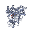
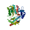
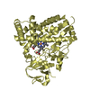
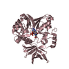
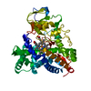
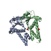
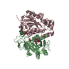
| Title | Crystal structure of LpqY from Mycobacterium thermoresistible in complex with trehalose | |||||||||||||||
 Components Components | Lipoprotein (Sugar-binding) lpqY | |||||||||||||||
 Keywords Keywords | TRANSPORT PROTEIN / LpqY / ABC-transporter / trehalose complex / tuberculosis | |||||||||||||||
| Function / homology | : / Bacterial extracellular solute-binding protein / Bacterial extracellular solute-binding protein / Periplasmic binding protein-like II / D-Maltodextrin-Binding Protein; domain 2 / 3-Layer(aba) Sandwich / Alpha Beta / trehalose / Lipoprotein (Sugar-binding) lpqY Function and homology information Function and homology information | |||||||||||||||
| Biological species |  Mycolicibacterium thermoresistibile (bacteria) Mycolicibacterium thermoresistibile (bacteria) | |||||||||||||||
| Method |  X-RAY DIFFRACTION / SYNCHROTRON / MOLECULAR REPLACEMENT / Resolution: 1.7 Å X-RAY DIFFRACTION / SYNCHROTRON / MOLECULAR REPLACEMENT / Resolution: 1.7 Å | |||||||||||||||
 Authors Authors | Furze, C.M. / Guy, C.M. / Angula, J. / Cameron, A.D. / Fullam, E. | |||||||||||||||
| Funding support |  United Kingdom, 4items United Kingdom, 4items
| |||||||||||||||
 Citation Citation | Journal: J.Biol.Chem. / Year: 2021 Title: Structural basis of trehalose recognition by the mycobacterial LpqY-SugABC transporter. Authors: Furze, C.M. / Delso, I. / Casal, E. / Guy, C.S. / Seddon, C. / Brown, C.M. / Parker, H.L. / Radhakrishnan, A. / Pacheco-Gomez, R. / Stansfeld, P.J. / Angulo, J. / Cameron, A.D. / Fullam, E. | |||||||||||||||
| History |
|
- Structure visualization
| Structure viewer | Molecule: MolmilJmol/JSmol |
|---|
- Downloads & links
-Download
| PDBx/mmCIF format | 7ape.cif.gz | 405.8 KB | Display | PDBx/mmCIF format |
|---|---|---|---|---|
| PDB format | pdb7ape.ent.gz | 268.8 KB | Display | PDB format |
| PDBx/mmJSON format | 7ape.json.gz | Tree view | PDBx/mmJSON format | |
| Others |  Other downloads Other downloads |
-Validation report
| Arichive directory | https://data.pdbj.org/pub/pdb/validation_reports/ap/7apeftp://data.pdbj.org/pub/pdb/validation_reports/ap/7ape | HTTPS FTP |
|---|
-Related structure data
| Similar structure data |
|---|


-Links
 PDBj
PDBj


- Assembly
| Deposited unit | 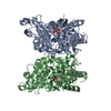
| ||||||||||||
|---|---|---|---|---|---|---|---|---|---|---|---|---|---|
| 1 | 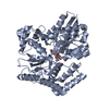
| ||||||||||||
| 2 | 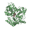
| ||||||||||||
| 3 |
| ||||||||||||
| Unit cell |
| ||||||||||||
| Components on special symmetry positions |
|
-Components
| #1: Protein | Mass: 48785.309 Da / Num. of mol.: 2 Source method: isolated from a genetically manipulated source Source: (gene. exp.) Mycolicibacterium thermoresistibile (bacteria)Gene: RMCT_2827 / Production host: #2: Polysaccharide | 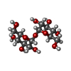  Source method: isolated from a genetically manipulated source Details: oligosaccharide with reducing-end-to-reducing-end glycosidic bond References: trehalose #3: Water | ChemComp-HOH / |  Mass: 18.015 Da / Num. of mol.: 557 / Source method: isolated from a natural source / Formula: H2O Mass: 18.015 Da / Num. of mol.: 557 / Source method: isolated from a natural source / Formula: H2OHas ligand of interest | Y | Has protein modification | Y | |
|---|
-Experimental details
-Experiment
| Experiment | Method: X-RAY DIFFRACTION / Number of used crystals: 1 |
|---|
- Sample preparation
| Crystal | Density Matthews: 2.4 Å3/Da / Density % sol: 48.84 % |
|---|---|
| Crystal grow | Temperature: 292 K / Method: vapor diffusion, sitting drop / pH: 6 Details: 0.1 M Hepes pH 6.0, 50 % w/v polypropylene glycol 400, 5 % DMSO, 1 mM TCEP |
-Data collection
| Diffraction | Mean temperature: 80 K / Serial crystal experiment: N |
|---|---|
| Diffraction source | Source: SYNCHROTRON / Site: Diamond / Beamline: I03 / Wavelength: 0.97 Å |
| Detector | Type: DECTRIS EIGER2 XE 16M / Detector: PIXEL / Date: May 11, 2019 |
| Radiation | Protocol: SINGLE WAVELENGTH / Monochromatic (M) / Laue (L): M / Scattering type: x-ray |
| Radiation wavelength | Wavelength: 0.97 Å / Relative weight: 1 |
| Reflection | Resolution: 1.7→54.1 Å / Num. obs: 102435 / % possible obs: 98.7 % / Redundancy: 23 % / Biso Wilson estimate: 26.14 Å2 / CC1/2: 0.999 / Net I/σ(I): 15.16 |
| Reflection shell | Resolution: 1.7→1.76 Å / Mean I/σ(I) obs: 1.2 / Num. unique obs: 9278 / Rpim(I) all: 0.57 |
- Processing
| Software |
| |||||||||||||||||||||||||||||||||||||||||||||||||||||||||||||||||||||||||||||||||||||||||||||||||||||||||||||||||||||||||||||||||||||||||||||||||||||||||||||||||||||||||||||||||||||||||||||||||||||||||||
|---|---|---|---|---|---|---|---|---|---|---|---|---|---|---|---|---|---|---|---|---|---|---|---|---|---|---|---|---|---|---|---|---|---|---|---|---|---|---|---|---|---|---|---|---|---|---|---|---|---|---|---|---|---|---|---|---|---|---|---|---|---|---|---|---|---|---|---|---|---|---|---|---|---|---|---|---|---|---|---|---|---|---|---|---|---|---|---|---|---|---|---|---|---|---|---|---|---|---|---|---|---|---|---|---|---|---|---|---|---|---|---|---|---|---|---|---|---|---|---|---|---|---|---|---|---|---|---|---|---|---|---|---|---|---|---|---|---|---|---|---|---|---|---|---|---|---|---|---|---|---|---|---|---|---|---|---|---|---|---|---|---|---|---|---|---|---|---|---|---|---|---|---|---|---|---|---|---|---|---|---|---|---|---|---|---|---|---|---|---|---|---|---|---|---|---|---|---|---|---|---|---|---|---|---|
| Refinement | Method to determine structure: MOLECULAR REPLACEMENT Starting model: LpqY iodide soak Resolution: 1.7→48.4 Å / SU ML: 0.2054 / Cross valid method: FREE R-VALUE / σ(F): 1.34 / Phase error: 19.5685 Stereochemistry target values: GeoStd + Monomer Library + CDL v1.2
| |||||||||||||||||||||||||||||||||||||||||||||||||||||||||||||||||||||||||||||||||||||||||||||||||||||||||||||||||||||||||||||||||||||||||||||||||||||||||||||||||||||||||||||||||||||||||||||||||||||||||||
| Solvent computation | Shrinkage radii: 0.9 Å / VDW probe radii: 1.11 Å / Solvent model: FLAT BULK SOLVENT MODEL | |||||||||||||||||||||||||||||||||||||||||||||||||||||||||||||||||||||||||||||||||||||||||||||||||||||||||||||||||||||||||||||||||||||||||||||||||||||||||||||||||||||||||||||||||||||||||||||||||||||||||||
| Displacement parameters | Biso mean: 29.75 Å2 | |||||||||||||||||||||||||||||||||||||||||||||||||||||||||||||||||||||||||||||||||||||||||||||||||||||||||||||||||||||||||||||||||||||||||||||||||||||||||||||||||||||||||||||||||||||||||||||||||||||||||||
| Refinement step | Cycle: LAST / Resolution: 1.7→48.4 Å
| |||||||||||||||||||||||||||||||||||||||||||||||||||||||||||||||||||||||||||||||||||||||||||||||||||||||||||||||||||||||||||||||||||||||||||||||||||||||||||||||||||||||||||||||||||||||||||||||||||||||||||
| Refine LS restraints |
| |||||||||||||||||||||||||||||||||||||||||||||||||||||||||||||||||||||||||||||||||||||||||||||||||||||||||||||||||||||||||||||||||||||||||||||||||||||||||||||||||||||||||||||||||||||||||||||||||||||||||||
| LS refinement shell |
|
