 Movie
Movie Controller

[English] 日本語
 Yorodumi
- PDB-2yve: Crystal structure of the methylene blue-bound form of the multi-d... -
+ Open data

- Basic information
| Entry | Database: PDB / ID: 2yve | ||||||
|---|---|---|---|---|---|---|---|
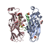
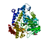
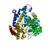
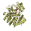
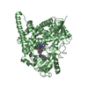
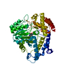
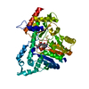
| Title | Crystal structure of the methylene blue-bound form of the multi-drug binding transcriptional repressor CgmR | ||||||
 Components Components | Transcriptional regulator | ||||||
 Keywords Keywords | TRANSCRIPTION / transcriptional regulator / helix-turn-helix / TetR-family | ||||||
| Function / homology |  Function and homology information Function and homology information | ||||||
| Biological species |  Corynebacterium glutamicum (bacteria) Corynebacterium glutamicum (bacteria) | ||||||
| Method |  X-RAY DIFFRACTION / SYNCHROTRON / MOLECULAR REPLACEMENT / Resolution: 1.4 Å X-RAY DIFFRACTION / SYNCHROTRON / MOLECULAR REPLACEMENT / Resolution: 1.4 Å | ||||||
 Authors Authors | Itou, H. / Shirakihara, Y. / Tanaka, I. | ||||||
 Citation Citation | Journal: J.Mol.Biol. / Year: 2010 Title: Crystal Structures of the Multidrug Binding Repressor Corynebacteriumglutamicum CgmR in Complex with Inducers and with an Operator Authors: Itou, H. / Watanabe, N. / Yao, M. / Shirakihara, Y. / Tanaka, I. | ||||||
| History |
|
- Structure visualization
| Structure viewer | Molecule: MolmilJmol/JSmol |
|---|
- Downloads & links
-Download
| PDBx/mmCIF format | 2yve.cif.gz | 91.5 KB | Display | PDBx/mmCIF format |
|---|---|---|---|---|
| PDB format | pdb2yve.ent.gz | 69 KB | Display | PDB format |
| PDBx/mmJSON format | 2yve.json.gz | Tree view | PDBx/mmJSON format | |
| Others |  Other downloads Other downloads |
-Validation report
| Arichive directory | https://data.pdbj.org/pub/pdb/validation_reports/yv/2yveftp://data.pdbj.org/pub/pdb/validation_reports/yv/2yve | HTTPS FTP |
|---|
-Related structure data
| Related structure data | 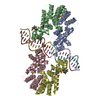 2yvhC  2zozC  1v7b C: citing same article ( S: Starting model for refinement |
|---|---|
| Similar structure data |

-Links
 PDBj
PDBj- Assembly
| Deposited unit | 
| ||||||||
|---|---|---|---|---|---|---|---|---|---|
| 1 |
| ||||||||
| Unit cell |
|
-Components
| #1: Protein | Mass: 21356.125 Da / Num. of mol.: 2 Source method: isolated from a genetically manipulated source Source: (gene. exp.) Corynebacterium glutamicum (bacteria) / Strain: ATCC13032 / Gene: cgl2612 / Plasmid: pET26b / Production host: #2: Chemical | ChemComp-CL / |   Mass: 35.453 Da / Num. of mol.: 1 / Source method: obtained synthetically / Formula: Cl Mass: 35.453 Da / Num. of mol.: 1 / Source method: obtained synthetically / Formula: Cl#3: Chemical | 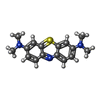  Mass: 284.399 Da / Num. of mol.: 2 / Source method: obtained synthetically / Formula: C16H18N3S / Comment: medication*YM Mass: 284.399 Da / Num. of mol.: 2 / Source method: obtained synthetically / Formula: C16H18N3S / Comment: medication*YM#4: Chemical | ChemComp-GOL /   Mass: 92.094 Da / Num. of mol.: 6 / Source method: obtained synthetically / Formula: C3H8O3 Mass: 92.094 Da / Num. of mol.: 6 / Source method: obtained synthetically / Formula: C3H8O3#5: Water | ChemComp-HOH / |  Mass: 18.015 Da / Num. of mol.: 322 / Source method: isolated from a natural source / Formula: H2O Mass: 18.015 Da / Num. of mol.: 322 / Source method: isolated from a natural source / Formula: H2O |
|---|
-Experimental details
-Experiment
| Experiment | Method: X-RAY DIFFRACTION / Number of used crystals: 1 |
|---|
- Sample preparation
| Crystal | Density Matthews: 2.41 Å3/Da / Density % sol: 49.04 % |
|---|---|
| Crystal grow | Temperature: 293 K / Method: vapor diffusion, hanging drop / pH: 6 Details: 0.1M Cacodylate, 0.1M MgCl2, 8% PEG 3350, pH 6.0, VAPOR DIFFUSION, HANGING DROP, temperature 293K |
-Data collection
| Diffraction | Mean temperature: 100 K |
|---|---|
| Diffraction source | Source: SYNCHROTRON / Site: Photon Factory  / Beamline: BL-17A / Wavelength: 1 Å / Beamline: BL-17A / Wavelength: 1 Å |
| Detector | Type: ADSC QUANTUM 4 / Detector: CCD / Date: Feb 16, 2007 / Details: mirrors |
| Radiation | Monochromator: Si(111) / Protocol: SINGLE WAVELENGTH / Monochromatic (M) / Laue (L): M / Scattering type: x-ray |
| Radiation wavelength | Wavelength: 1 Å / Relative weight: 1 |
| Reflection | Resolution: 1.4→50 Å / Num. all: 78066 / Num. obs: 78066 / % possible obs: 95.3 % / Observed criterion σ(F): -3 / Redundancy: 7.5 % / Biso Wilson estimate: 15 Å2 / Rmerge(I) obs: 0.052 / Net I/σ(I): 19.7 |
| Reflection shell | Resolution: 1.4→1.45 Å / Redundancy: 7.6 % / Rmerge(I) obs: 0.228 / Num. unique all: 7625 / % possible all: 94.5 |
- Processing
| Software |
| ||||||||||||||||||||||||||||||||||||
|---|---|---|---|---|---|---|---|---|---|---|---|---|---|---|---|---|---|---|---|---|---|---|---|---|---|---|---|---|---|---|---|---|---|---|---|---|---|
| Refinement | Method to determine structure: MOLECULAR REPLACEMENT Starting model: PDB entry 1V7B 1v7b Resolution: 1.4→9.97 Å / Rfactor Rfree error: 0.003 / Data cutoff high absF: 1296929.63 / Data cutoff low absF: 0 / Isotropic thermal model: RESTRAINED / Cross valid method: THROUGHOUT / σ(F): 0 / σ(I): 0 / Stereochemistry target values: Engh & Huber
| ||||||||||||||||||||||||||||||||||||
| Solvent computation | Solvent model: FLAT MODEL / Bsol: 76.5288 Å2 / ksol: 0.585223 e/Å3 | ||||||||||||||||||||||||||||||||||||
| Displacement parameters | Biso mean: 17.6 Å2
| ||||||||||||||||||||||||||||||||||||
| Refine analyze |
| ||||||||||||||||||||||||||||||||||||
| Refinement step | Cycle: LAST / Resolution: 1.4→9.97 Å
| ||||||||||||||||||||||||||||||||||||
| Refine LS restraints |
| ||||||||||||||||||||||||||||||||||||
| LS refinement shell | Resolution: 1.4→1.49 Å / Rfactor Rfree error: 0.007 / Total num. of bins used: 6
| ||||||||||||||||||||||||||||||||||||
| Xplor file |
|
