Movie
Movie Controller


+ Open data

- Basic information
| Entry | Database: PDB / ID: 7ai5 | ||||||
|---|---|---|---|---|---|---|---|
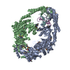
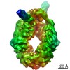
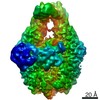
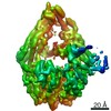
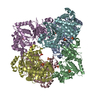
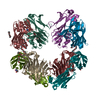
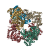
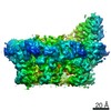
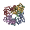
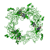
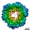
| Title | MutS in Scanning state | ||||||
 Components Components |
| ||||||
 Keywords Keywords | DNA BINDING PROTEIN / DNA Mismatch Repair MutS | ||||||
| Function / homology |  Function and homology information Function and homology informationmismatched DNA binding / ATP-dependent DNA damage sensor activity / mismatch repair / damaged DNA binding / ATP binding / cytosol Similarity search - Function | ||||||
| Biological species |  synthetic construct (others) | ||||||
| Method | ELECTRON MICROSCOPY / single particle reconstruction / cryo EM / Resolution: 4.4 Å | ||||||
 Authors Authors | Fernandez-Leiro, R. / Bhairosing-Kok, D. / Sixma, T.K. / Lamers, M.H. | ||||||
| Funding support |  United Kingdom, 1items United Kingdom, 1items
| ||||||
 Citation Citation | Journal: Nat Struct Mol Biol / Year: 2021 Title: The selection process of licensing a DNA mismatch for repair. Authors: Rafael Fernandez-Leiro / Doreth Bhairosing-Kok / Vladislav Kunetsky / Charlie Laffeber / Herrie H Winterwerp / Flora Groothuizen / Alexander Fish / Joyce H G Lebbink / Peter Friedhoff / ...Authors: Rafael Fernandez-Leiro / Doreth Bhairosing-Kok / Vladislav Kunetsky / Charlie Laffeber / Herrie H Winterwerp / Flora Groothuizen / Alexander Fish / Joyce H G Lebbink / Peter Friedhoff / Titia K Sixma / Meindert H Lamers /    Abstract: DNA mismatch repair detects and removes mismatches from DNA by a conserved mechanism, reducing the error rate of DNA replication by 100- to 1,000-fold. In this process, MutS homologs scan DNA, ...DNA mismatch repair detects and removes mismatches from DNA by a conserved mechanism, reducing the error rate of DNA replication by 100- to 1,000-fold. In this process, MutS homologs scan DNA, recognize mismatches and initiate repair. How the MutS homologs selectively license repair of a mismatch among millions of matched base pairs is not understood. Here we present four cryo-EM structures of Escherichia coli MutS that provide snapshots, from scanning homoduplex DNA to mismatch binding and MutL activation via an intermediate state. During scanning, the homoduplex DNA forms a steric block that prevents MutS from transitioning into the MutL-bound clamp state, which can only be overcome through kinking of the DNA at a mismatch. Structural asymmetry in all four structures indicates a division of labor between the two MutS monomers. Together, these structures reveal how a small conformational change from the homoduplex- to heteroduplex-bound MutS acts as a licensing step that triggers a dramatic conformational change that enables MutL binding and initiation of the repair cascade. | ||||||
| History |
|
- Structure visualization
| Movie |
Movie viewer |
|---|---|
| Structure viewer | Molecule: MolmilJmol/JSmol |
- Downloads & links
-Download
| PDBx/mmCIF format | 7ai5.cif.gz | 323.5 KB | Display | PDBx/mmCIF format |
|---|---|---|---|---|
| PDB format | pdb7ai5.ent.gz | 250.9 KB | Display | PDB format |
| PDBx/mmJSON format | 7ai5.json.gz | Tree view | PDBx/mmJSON format | |
| Others |  Other downloads Other downloads |
-Validation report
| Arichive directory | https://data.pdbj.org/pub/pdb/validation_reports/ai/7ai5ftp://data.pdbj.org/pub/pdb/validation_reports/ai/7ai5 | HTTPS FTP |
|---|
-Related structure data
| Related structure data | 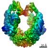 11791MC 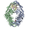 7ai6C 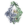 7ai7C 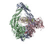 7aibC 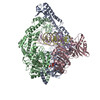 7aicC M: map data used to model this data C: citing same article ( |
|---|---|
| Similar structure data |

-Links
 PDBj
PDBj


- Assembly
| Deposited unit | 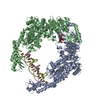
| ||||||||||||||||||
|---|---|---|---|---|---|---|---|---|---|---|---|---|---|---|---|---|---|---|---|
| 1 |
| ||||||||||||||||||
| Noncrystallographic symmetry (NCS) | NCS domain:
NCS domain segments: Component-ID: _ / Ens-ID: 1 / Beg auth comp-ID: SER / Beg label comp-ID: SER / End auth comp-ID: SER / End label comp-ID: SER / Refine code: _ / Auth seq-ID: 2 - 800 / Label seq-ID: 2 - 800
|
-Components
| #1: Protein | Mass: 95397.898 Da / Num. of mol.: 2 / Mutation: D835R Source method: isolated from a genetically manipulated source Source: (gene. exp.) Gene: mutS, ACU57_10490, AUQ13_17300, BANRA_01250, BANRA_04372, BMA87_05210, BvCms2454_03957, BvCmsKSP026_01123, BvCmsSINP011_00857, C9Z39_17590, CI693_21650, D2185_15190, D3821_16315, D4638_12960, ...Gene: mutS, ACU57_10490, AUQ13_17300, BANRA_01250, BANRA_04372, BMA87_05210, BvCms2454_03957, BvCmsKSP026_01123, BvCmsSINP011_00857, C9Z39_17590, CI693_21650, D2185_15190, D3821_16315, D4638_12960, D9D20_12730, D9D44_11420, DAH34_11930, DJ503_05760, DL326_19385, DT034_19915, DTL43_09115, E2119_10690, E2127_10890, E2128_08030, EAI52_13070, EC3234A_48c00420, ECTO6_01123, ED307_14565, EEP23_04760, EI041_14570, EL75_0954, EL79_0966, EL80_0968, ELT20_17315, EPT01_07440, FORC82_1112, FV293_17550, GHR40_09850, GKF74_12505, GKF86_14440, GKF89_15940, NCTC10963_01107, NCTC13216_00777, NCTC8500_01160, NCTC9045_01268, NCTC9062_02087, RK56_022910, SAMEA3484427_01823, SAMEA3484429_01685, SAMEA3752559_02626, SAMEA3753300_03556 Production host: #2: DNA chain | | Mass: 6832.414 Da / Num. of mol.: 1 / Source method: obtained synthetically Details: Plasmid DNA molecule (pRC1765), sequence in structure unidentified Source: (synth.) synthetic construct (others) #3: DNA chain | | Mass: 6672.318 Da / Num. of mol.: 1 / Source method: obtained synthetically Details: Plasmid DNA molecule (pRC1765), sequence in structure unidentified Source: (synth.) synthetic construct (others) #4: Chemical |   Mass: 507.181 Da / Num. of mol.: 2 / Source method: obtained synthetically / Formula: C10H16N5O13P3 / Comment: ATP, energy-carrying molecule*YM Mass: 507.181 Da / Num. of mol.: 2 / Source method: obtained synthetically / Formula: C10H16N5O13P3 / Comment: ATP, energy-carrying molecule*YMHas ligand of interest | N | |
|---|
-Experimental details
-Experiment
| Experiment | Method: ELECTRON MICROSCOPY |
|---|---|
| EM experiment | Aggregation state: PARTICLE / 3D reconstruction method: single particle reconstruction |
- Sample preparation
| Component |
| ||||||||||||||||||||||||
|---|---|---|---|---|---|---|---|---|---|---|---|---|---|---|---|---|---|---|---|---|---|---|---|---|---|
| Molecular weight | Value: 0.190 MDa / Experimental value: NO | ||||||||||||||||||||||||
| Source (natural) |
| ||||||||||||||||||||||||
| Source (recombinant) |
| ||||||||||||||||||||||||
| Buffer solution | pH: 7.5 | ||||||||||||||||||||||||
| Buffer component |
| ||||||||||||||||||||||||
| Specimen | Conc.: 1 mg/ml / Embedding applied: NO / Shadowing applied: NO / Staining applied: NO / Vitrification applied: YES Details: Protein sample was purified over a gel filtration column and mixed with DNA+ATP prior to grid preparation | ||||||||||||||||||||||||
| Specimen support | Grid material: COPPER / Grid mesh size: 300 divisions/in. / Grid type: Quantifoil | ||||||||||||||||||||||||
| Vitrification | Instrument: FEI VITROBOT MARK IV / Cryogen name: ETHANE / Humidity: 100 % / Chamber temperature: 277 K / Details: blot for 3 seconds before plunging |
- Electron microscopy imaging
| Experimental equipment |  Model: Titan Krios / Image courtesy: FEI Company |
|---|---|
| Microscopy | Model: FEI TITAN KRIOS |
| Electron gun | Electron source:  FIELD EMISSION GUN / Accelerating voltage: 300 kV / Illumination mode: FLOOD BEAM FIELD EMISSION GUN / Accelerating voltage: 300 kV / Illumination mode: FLOOD BEAM |
| Electron lens | Mode: BRIGHT FIELD / Nominal magnification: 105000 X / Calibrated defocus min: 500 nm / Calibrated defocus max: 4000 nm / Cs: 2.7 mm / C2 aperture diameter: 100 µm / Alignment procedure: COMA FREE |
| Specimen holder | Cryogen: NITROGEN / Specimen holder model: FEI TITAN KRIOS AUTOGRID HOLDER |
| Image recording | Average exposure time: 12 sec. / Electron dose: 40 e/Å2 / Detector mode: COUNTING / Film or detector model: GATAN K2 SUMMIT (4k x 4k) / Num. of grids imaged: 1 / Num. of real images: 2351 |
| EM imaging optics | Energyfilter name: GIF Quantum LS / Energyfilter slit width: 20 eV |
| Image scans | Sampling size: 5 µm / Width: 3838 / Height: 3710 / Movie frames/image: 40 / Used frames/image: 1-40 |
- Processing
| Software | Name: REFMAC / Version: 5.8.0258 / Classification: refinement | ||||||||||||||||||||||||||||||||||||||||||||||||||||||||||||||||||||||||||||||||||||||||||||||||||||||||||
|---|---|---|---|---|---|---|---|---|---|---|---|---|---|---|---|---|---|---|---|---|---|---|---|---|---|---|---|---|---|---|---|---|---|---|---|---|---|---|---|---|---|---|---|---|---|---|---|---|---|---|---|---|---|---|---|---|---|---|---|---|---|---|---|---|---|---|---|---|---|---|---|---|---|---|---|---|---|---|---|---|---|---|---|---|---|---|---|---|---|---|---|---|---|---|---|---|---|---|---|---|---|---|---|---|---|---|---|
| EM software |
| ||||||||||||||||||||||||||||||||||||||||||||||||||||||||||||||||||||||||||||||||||||||||||||||||||||||||||
| CTF correction | Type: PHASE FLIPPING AND AMPLITUDE CORRECTION | ||||||||||||||||||||||||||||||||||||||||||||||||||||||||||||||||||||||||||||||||||||||||||||||||||||||||||
| Particle selection | Num. of particles selected: 229664 | ||||||||||||||||||||||||||||||||||||||||||||||||||||||||||||||||||||||||||||||||||||||||||||||||||||||||||
| Symmetry | Point symmetry: C1 (asymmetric) | ||||||||||||||||||||||||||||||||||||||||||||||||||||||||||||||||||||||||||||||||||||||||||||||||||||||||||
| 3D reconstruction | Resolution: 4.4 Å / Resolution method: FSC 0.143 CUT-OFF / Num. of particles: 36682 / Algorithm: BACK PROJECTION / Symmetry type: POINT | ||||||||||||||||||||||||||||||||||||||||||||||||||||||||||||||||||||||||||||||||||||||||||||||||||||||||||
| Atomic model building | Protocol: FLEXIBLE FIT / Space: RECIPROCAL Details: Initial Jelly Body refinement Final refinement with proSmart restraints | ||||||||||||||||||||||||||||||||||||||||||||||||||||||||||||||||||||||||||||||||||||||||||||||||||||||||||
| Atomic model building | PDB-ID: 1E3M Accession code: 1E3M / Source name: PDB / Type: experimental model | ||||||||||||||||||||||||||||||||||||||||||||||||||||||||||||||||||||||||||||||||||||||||||||||||||||||||||
| Refinement | Resolution: 4.4→4.4 Å / Cor.coef. Fo:Fc: 0.944 / SU B: 186.237 / SU ML: 2.008 Stereochemistry target values: MAXIMUM LIKELIHOOD WITH PHASES Details: HYDROGENS HAVE BEEN ADDED IN THE RIDING POSITIONS
| ||||||||||||||||||||||||||||||||||||||||||||||||||||||||||||||||||||||||||||||||||||||||||||||||||||||||||
| Solvent computation | Solvent model: PARAMETERS FOR MASK CACLULATION | ||||||||||||||||||||||||||||||||||||||||||||||||||||||||||||||||||||||||||||||||||||||||||||||||||||||||||
| Displacement parameters | Biso mean: 117.105 Å2
| ||||||||||||||||||||||||||||||||||||||||||||||||||||||||||||||||||||||||||||||||||||||||||||||||||||||||||
| Refinement step | Cycle: 1 / Total: 13316 | ||||||||||||||||||||||||||||||||||||||||||||||||||||||||||||||||||||||||||||||||||||||||||||||||||||||||||
| Refine LS restraints |
|