 Movie
Movie Controller

[English] 日本語
 Yorodumi
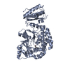
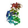
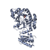
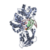
- PDB-7agp: Structure of the AcylTransferase domain of Mycocerosic Acid Synth... -
+ Open data

- Basic information
| Entry | Database: PDB / ID: 7agp | ||||||
|---|---|---|---|---|---|---|---|
| Title | Structure of the AcylTransferase domain of Mycocerosic Acid Synthase from Mycobacterium tuberculosis | ||||||
 Components Components | Mycocerosic acid synthase | ||||||
 Keywords Keywords | TRANSFERASE / Mycocerosic Acid Synthase / AcylTransferase domain / polyketide synthase / Mycobacterium tuberculosis | ||||||
| Function / homology |  Function and homology information Function and homology informationmycocerosate synthase / mycocerosate synthase activity / DIM/DIP cell wall layer assembly / fatty acid synthase activity / ligase activity / phosphopantetheine binding / 3-oxoacyl-[acyl-carrier-protein] synthase activity / fatty acid biosynthetic process / oxidoreductase activity / hydrolase activity ...mycocerosate synthase / mycocerosate synthase activity / DIM/DIP cell wall layer assembly / fatty acid synthase activity / ligase activity / phosphopantetheine binding / 3-oxoacyl-[acyl-carrier-protein] synthase activity / fatty acid biosynthetic process / oxidoreductase activity / hydrolase activity / plasma membrane / cytoplasm Similarity search - Function | ||||||
| Biological species |  Mycobacterium bovis (bacteria) Mycobacterium bovis (bacteria) | ||||||
| Method |  X-RAY DIFFRACTION / SYNCHROTRON / MOLECULAR REPLACEMENT / Resolution: 2.4 Å X-RAY DIFFRACTION / SYNCHROTRON / MOLECULAR REPLACEMENT / Resolution: 2.4 Å | ||||||
 Authors Authors | Brison, Y. / Nahoum, V. / Mourey, L. / Maveyraud, L. | ||||||
| Funding support |  France, 1items France, 1items
| ||||||
 Citation Citation | Journal: Acs Chem.Biol. / Year: 2020 Title: Molecular Basis for Extender Unit Specificity of Mycobacterial Polyketide Synthases. Authors: Grabowska, A.D. / Brison, Y. / Maveyraud, L. / Gavalda, S. / Faille, A. / Nahoum, V. / Bon, C. / Guilhot, C. / Pedelacq, J.D. / Chalut, C. / Mourey, L. | ||||||
| History |
|
- Structure visualization
| Structure viewer | Molecule: MolmilJmol/JSmol |
|---|
- Downloads & links
-Download
| PDBx/mmCIF format | 7agp.cif.gz | 330.8 KB | Display | PDBx/mmCIF format |
|---|---|---|---|---|
| PDB format | pdb7agp.ent.gz | 271.6 KB | Display | PDB format |
| PDBx/mmJSON format | 7agp.json.gz | Tree view | PDBx/mmJSON format | |
| Others |  Other downloads Other downloads |
-Validation report
| Arichive directory | https://data.pdbj.org/pub/pdb/validation_reports/ag/7agpftp://data.pdbj.org/pub/pdb/validation_reports/ag/7agp | HTTPS FTP |
|---|
-Related structure data
| Related structure data | 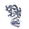 7agqC 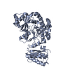 7agrC 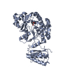 7agsC  7agtC  7aguC  7ahbC 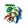 7akcC  3tzwS C: citing same article ( S: Starting model for refinement |
|---|---|
| Similar structure data |


-Links
 PDBj
PDBj


- Assembly
| Deposited unit | 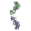
| ||||||||
|---|---|---|---|---|---|---|---|---|---|
| 1 | 
| ||||||||
| 2 | 
| ||||||||
| Unit cell |
|
-Components
| #1: Protein | Mass: 46470.418 Da / Num. of mol.: 2 Source method: isolated from a genetically manipulated source Source: (gene. exp.) Mycobacterium bovis (strain ATCC BAA-935 / AF2122/97) (bacteria)Strain: ATCC BAA-935 / AF2122/97 / Gene: mas, BQ2027_MB2965C / Production host: #2: Chemical |   Mass: 62.068 Da / Num. of mol.: 2 / Source method: obtained synthetically / Formula: C2H6O2 Mass: 62.068 Da / Num. of mol.: 2 / Source method: obtained synthetically / Formula: C2H6O2#3: Water | ChemComp-HOH / |  Mass: 18.015 Da / Num. of mol.: 238 / Source method: isolated from a natural source / Formula: H2O Mass: 18.015 Da / Num. of mol.: 238 / Source method: isolated from a natural source / Formula: H2OHas ligand of interest | N | Has protein modification | Y | |
|---|
-Experimental details
-Experiment
| Experiment | Method: X-RAY DIFFRACTION / Number of used crystals: 1 |
|---|
- Sample preparation
| Crystal | Density Matthews: 3.53 Å3/Da / Density % sol: 65.12 % |
|---|---|
| Crystal grow | Temperature: 293 K / Method: vapor diffusion, sitting drop / pH: 4.2 / Details: 1.1 M sodium succinate 0.1 M sodium acetate buffer |
-Data collection
| Diffraction | Mean temperature: 100 K / Serial crystal experiment: N |
|---|---|
| Diffraction source | Source: SYNCHROTRON / Site: ESRF / Beamline: ID23-1 / Wavelength: 0.9763 Å |
| Detector | Type: ADSC QUANTUM 315 / Detector: CCD / Date: Sep 30, 2011 |
| Radiation | Protocol: SINGLE WAVELENGTH / Monochromatic (M) / Laue (L): M / Scattering type: x-ray |
| Radiation wavelength | Wavelength: 0.9763 Å / Relative weight: 1 |
| Reflection | Resolution: 2.4→49.3 Å / Num. obs: 51153 / % possible obs: 99.1 % / Redundancy: 3.54 % / Biso Wilson estimate: 74.1 Å2 / CC1/2: 0.999 / Rmerge(I) obs: 0.042 / Rrim(I) all: 0.05 / Rsym value: 0.042 / Net I/σ(I): 15.77 |
| Reflection shell | Resolution: 2.4→2.54 Å / Redundancy: 3.48 % / Rmerge(I) obs: 0.845 / Mean I/σ(I) obs: 1.43 / Num. unique obs: 8121 / CC1/2: 0.658 / Rrim(I) all: 0.998 / Rsym value: 0.845 / % possible all: 98.4 |
- Processing
| Software |
| |||||||||||||||||||||||||||||||||||||||||||||||||||||||||||||||||||||||||||
|---|---|---|---|---|---|---|---|---|---|---|---|---|---|---|---|---|---|---|---|---|---|---|---|---|---|---|---|---|---|---|---|---|---|---|---|---|---|---|---|---|---|---|---|---|---|---|---|---|---|---|---|---|---|---|---|---|---|---|---|---|---|---|---|---|---|---|---|---|---|---|---|---|---|---|---|---|
| Refinement | Method to determine structure: MOLECULAR REPLACEMENT Starting model: 3TZW Resolution: 2.4→49.3 Å / Cor.coef. Fo:Fc: 0.952 / Cor.coef. Fo:Fc free: 0.944 / SU R Cruickshank DPI: 0.243 / Cross valid method: THROUGHOUT / SU R Blow DPI: 0.243 / SU Rfree Blow DPI: 0.187 / SU Rfree Cruickshank DPI: 0.189
| |||||||||||||||||||||||||||||||||||||||||||||||||||||||||||||||||||||||||||
| Displacement parameters | Biso mean: 85.1 Å2
| |||||||||||||||||||||||||||||||||||||||||||||||||||||||||||||||||||||||||||
| Refine analyze | Luzzati coordinate error obs: 0.33 Å | |||||||||||||||||||||||||||||||||||||||||||||||||||||||||||||||||||||||||||
| Refinement step | Cycle: LAST / Resolution: 2.4→49.3 Å
| |||||||||||||||||||||||||||||||||||||||||||||||||||||||||||||||||||||||||||
| Refine LS restraints |
| |||||||||||||||||||||||||||||||||||||||||||||||||||||||||||||||||||||||||||
| LS refinement shell | Resolution: 2.4→2.42 Å
| |||||||||||||||||||||||||||||||||||||||||||||||||||||||||||||||||||||||||||
| Refinement TLS params. | Refine-ID: X-RAY DIFFRACTION
| |||||||||||||||||||||||||||||||||||||||||||||||||||||||||||||||||||||||||||
| Refinement TLS group |
|
