 Movie
Movie Controller


+ Open data

- Basic information
| Entry | Database: PDB / ID: 7adj | ||||||
|---|---|---|---|---|---|---|---|
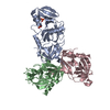
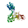
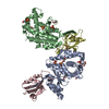
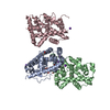
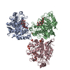
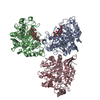
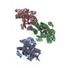
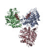
| Title | Structure of the mycoplasma MIB protein | ||||||
 Components Components | Putative immunoglobulin-blocking virulence protein | ||||||
 Keywords Keywords | MEMBRANE PROTEIN / Antibody binding protein / protease / protein complex. | ||||||
| Function / homology | Mycoplasma virulence, signal domain / Putative immunoglobulin-blocking virulence protein / Mycoplasma virulence signal region (Myco_arth_vir_N) / IgG-blocking virulence domain / Putative immunoglobulin-blocking virulence protein Function and homology information Function and homology information | ||||||
| Biological species |  Mycoplasma mycoides subsp. capri (bacteria) Mycoplasma mycoides subsp. capri (bacteria) | ||||||
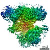
| Method | ELECTRON MICROSCOPY / single particle reconstruction / cryo EM / Resolution: 2.8 Å | ||||||
 Authors Authors | Nottelet, P. / Bataille, L. / Gourgues, G. / Anger, R. / Lartigue, C. / Sirand-Pugnet, P. / Marza, E. / Fronzes, R. / Arfi, Y. | ||||||
| Funding support |  France, 1items France, 1items
| ||||||
 Citation Citation | Journal: Sci Adv / Year: 2021 Title: The mycoplasma surface proteins MIB and MIP promote the dissociation of the antibody-antigen interaction. Authors: Pierre Nottelet / Laure Bataille / Geraldine Gourgues / Robin Anger / Carole Lartigue / Pascal Sirand-Pugnet / Esther Marza / Remi Fronzes / Yonathan Arfi / Abstract: Mycoplasma immunoglobulin binding (MIB) and mycoplasma immunoglobulin protease (MIP) are surface proteins found in the majority of mycoplasma species, acting sequentially to capture antibodies and ...Mycoplasma immunoglobulin binding (MIB) and mycoplasma immunoglobulin protease (MIP) are surface proteins found in the majority of mycoplasma species, acting sequentially to capture antibodies and cleave off their V domains. Cryo-electron microscopy structures show how MIB and MIP bind to a Fab fragment in a "hug of death" mechanism. As a result, the orientation of the V and V domains is twisted out of alignment, disrupting the antigen binding site. We also show that MIB-MIP has the ability to promote the dissociation of the antibody-antigen complex. This system is functional in cells and protects mycoplasmas from antibody-mediated agglutination. These results highlight the key role of the MIB-MIP system in immunity evasion by mycoplasmas through an unprecedented mechanism, and open exciting perspectives to use these proteins as potential tools in the antibody field. | ||||||
| History |
|
- Structure visualization
| Movie |
Movie viewer |
|---|---|
| Structure viewer | Molecule: MolmilJmol/JSmol |
- Downloads & links
-Download
| PDBx/mmCIF format | 7adj.cif.gz | 215.8 KB | Display | PDBx/mmCIF format |
|---|---|---|---|---|
| PDB format | pdb7adj.ent.gz | 171.2 KB | Display | PDB format |
| PDBx/mmJSON format | 7adj.json.gz | Tree view | PDBx/mmJSON format | |
| Others |  Other downloads Other downloads |
-Validation report
| Arichive directory | https://data.pdbj.org/pub/pdb/validation_reports/ad/7adjftp://data.pdbj.org/pub/pdb/validation_reports/ad/7adj | HTTPS FTP |
|---|
-Related structure data
| Related structure data | 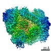 11727MC 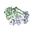 7adkC 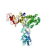 7admC M: map data used to model this data C: citing same article ( |
|---|---|
| Similar structure data |

-Links
 PDBj
PDBj- Assembly
| Deposited unit | 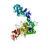
|
|---|---|
| 1 |
|
-Components
| #1: Protein | Mass: 82922.141 Da / Num. of mol.: 1 Source method: isolated from a genetically manipulated source Source: (gene. exp.) Mycoplasma mycoides subsp. capri (bacteria)Gene: MMC68F_00609 / Production host: |
|---|
-Experimental details
-Experiment
| Experiment | Method: ELECTRON MICROSCOPY |
|---|---|
| EM experiment | Aggregation state: PARTICLE / 3D reconstruction method: single particle reconstruction |
- Sample preparation
| Component | Name: Mycoplasma MIB-MIP proteins in complex with a goat Fab Type: COMPLEX / Entity ID: all / Source: RECOMBINANT |
|---|---|
| Molecular weight | Experimental value: NO |
| Source (natural) | Organism: Mycoplasma mycoides subsp. capri str. GM12 (bacteria) |
| Source (recombinant) | Organism: |
| Buffer solution | pH: 8 |
| Specimen | Embedding applied: NO / Shadowing applied: NO / Staining applied: NO / Vitrification applied: YES |
| Specimen support | Grid material: COPPER / Grid type: C-flat |
| Vitrification | Instrument: FEI VITROBOT MARK IV / Cryogen name: ETHANE / Humidity: 100 % / Chamber temperature: 4 K |
- Electron microscopy imaging
| Experimental equipment |  Model: Titan Krios / Image courtesy: FEI Company |
|---|---|
| Microscopy | Model: TFS KRIOS |
| Electron gun | Electron source:  FIELD EMISSION GUN / Accelerating voltage: 300 kV / Illumination mode: FLOOD BEAM FIELD EMISSION GUN / Accelerating voltage: 300 kV / Illumination mode: FLOOD BEAM |
| Electron lens | Mode: BRIGHT FIELD / Cs: 2.7 mm |
| Specimen holder | Cryogen: NITROGEN / Specimen holder model: FEI TITAN KRIOS AUTOGRID HOLDER |
| Image recording | Electron dose: 1.45 e/Å2 / Detector mode: COUNTING / Film or detector model: GATAN K2 SUMMIT (4k x 4k) |
| Image scans | Movie frames/image: 40 |
- Processing
| Software | Name: PHENIX / Version: 1.16_3546: / Classification: refinement | ||||||||||||||||||||||||
|---|---|---|---|---|---|---|---|---|---|---|---|---|---|---|---|---|---|---|---|---|---|---|---|---|---|
| EM software |
| ||||||||||||||||||||||||
| CTF correction | Type: PHASE FLIPPING AND AMPLITUDE CORRECTION | ||||||||||||||||||||||||
| 3D reconstruction | Resolution: 2.8 Å / Resolution method: FSC 0.143 CUT-OFF / Num. of particles: 255911 / Symmetry type: POINT | ||||||||||||||||||||||||
| Refine LS restraints |
|
