- PDB-7adm: Structure of the mycoplasma MIB protein -
+
Open data
ID or keywords:
Loading...
-
Basic information
Entry
Database: PDB / ID: 7adm
Title
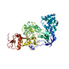
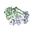
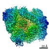
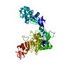
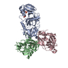
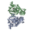
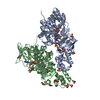
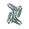
Structure of the mycoplasma MIB protein
Components
Putative immunoglobulin-blocking virulence protein
Keywords
MEMBRANE PROTEIN / Antibody binding protein / protease / protein complex.
Function / homology
Mycoplasma virulence, signal domain / Putative immunoglobulin-blocking virulence protein / Mycoplasma virulence signal region (Myco_arth_vir_N) / IgG-blocking virulence domain / Putative immunoglobulin-blocking virulence protein
Function and homology information
Biological species
Mycoplasma mycoides subsp. capri (bacteria)
Method
ELECTRON MICROSCOPY / single particle reconstruction / cryo EM / Resolution: 3.5 Å
Journal: Sci Adv / Year: 2021 Title: The mycoplasma surface proteins MIB and MIP promote the dissociation of the antibody-antigen interaction. Authors: Pierre Nottelet / Laure Bataille / Geraldine Gourgues / Robin Anger / Carole Lartigue / Pascal Sirand-Pugnet / Esther Marza / Remi Fronzes / Yonathan Arfi / Abstract: Mycoplasma immunoglobulin binding (MIB) and mycoplasma immunoglobulin protease (MIP) are surface proteins found in the majority of mycoplasma species, acting sequentially to capture antibodies and ...Mycoplasma immunoglobulin binding (MIB) and mycoplasma immunoglobulin protease (MIP) are surface proteins found in the majority of mycoplasma species, acting sequentially to capture antibodies and cleave off their V domains. Cryo-electron microscopy structures show how MIB and MIP bind to a Fab fragment in a "hug of death" mechanism. As a result, the orientation of the V and V domains is twisted out of alignment, disrupting the antigen binding site. We also show that MIB-MIP has the ability to promote the dissociation of the antibody-antigen complex. This system is functional in cells and protects mycoplasmas from antibody-mediated agglutination. These results highlight the key role of the MIB-MIP system in immunity evasion by mycoplasmas through an unprecedented mechanism, and open exciting perspectives to use these proteins as potential tools in the antibody field.
In the structure databanks used in Yorodumi, some data are registered as the other names, "COVID-19 virus" and "2019-nCoV". Here are the details of the virus and the list of structure data.
Jan 31, 2019. EMDB accession codes are about to change! (news from PDBe EMDB page)
EMDB accession codes are about to change! (news from PDBe EMDB page)
The allocation of 4 digits for EMDB accession codes will soon come to an end. Whilst these codes will remain in use, new EMDB accession codes will include an additional digit and will expand incrementally as the available range of codes is exhausted. The current 4-digit format prefixed with “EMD-” (i.e. EMD-XXXX) will advance to a 5-digit format (i.e. EMD-XXXXX), and so on. It is currently estimated that the 4-digit codes will be depleted around Spring 2019, at which point the 5-digit format will come into force.
The EM Navigator/Yorodumi systems omit the EMD- prefix.
Related info.:Q: What is EMD? / ID/Accession-code notation in Yorodumi/EM Navigator
Yorodumi is a browser for structure data from EMDB, PDB, SASBDB, etc.
This page is also the successor to EM Navigator detail page, and also detail information page/front-end page for Omokage search.
The word "yorodu" (or yorozu) is an old Japanese word meaning "ten thousand". "mi" (miru) is to see.
Related info.:EMDB / PDB / SASBDB / Comparison of 3 databanks / Yorodumi Search / Aug 31, 2016. New EM Navigator & Yorodumi / Yorodumi Papers / Jmol/JSmol / Function and homology information / Changes in new EM Navigator and Yorodumi
 Movie
Movie Controller


 Open data
 Basic information
 Components
 Keywords
 Function and homology information
 Mycoplasma mycoides subsp. capri (bacteria)
 Authors
 France, 1items
  Citation
 Structure visualization
 Downloads & links
 Other downloads
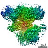


 PDBj
PDBj Assembly
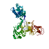
 Sample preparation
 Electron microscopy imaging
 FIELD EMISSION GUN / Accelerating voltage: 200 kV / Illumination mode: FLOOD BEAM
 Processing