 Movie
Movie Controller

[English] 日本語
 Yorodumi
- PDB-7ac6: Structure of sponge-phase grown PepTst2 collected by rotation ser... -
+ Open data

- Basic information
| Entry | Database: PDB / ID: 7ac6 | ||||||
|---|---|---|---|---|---|---|---|
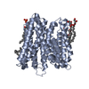
| Title | Structure of sponge-phase grown PepTst2 collected by rotation serial crystallography on a COC membrane at a synchrotron source | ||||||
 Components Components | (Di-or tripeptide:H+ ...) x 2 | ||||||
 Keywords Keywords | TRANSPORT PROTEIN / peptide transport protein | ||||||
| Function / homology |  Function and homology information Function and homology informationtripeptide transmembrane transport / tripeptide transmembrane transporter activity / peptide:proton symporter activity / dipeptide transmembrane transporter activity / identical protein binding / plasma membrane Similarity search - Function | ||||||
| Biological species |  Streptococcus thermophilus (bacteria) Streptococcus thermophilus (bacteria) | ||||||
| Method |  X-RAY DIFFRACTION / SYNCHROTRON / MOLECULAR REPLACEMENT / Resolution: 3 Å X-RAY DIFFRACTION / SYNCHROTRON / MOLECULAR REPLACEMENT / Resolution: 3 Å | ||||||
 Authors Authors | Martiel, I. / Padeste, C. / Karpik, A. / Huang, C.Y. / Wang, M. / Marsh, M. | ||||||
 Citation Citation | Journal: Acta Crystallogr D Struct Biol / Year: 2021 Title: Versatile microporous polymer-based supports for serial macromolecular crystallography. Authors: Martiel, I. / Beale, J.H. / Karpik, A. / Huang, C.Y. / Vera, L. / Olieric, N. / Wranik, M. / Tsai, C.J. / Muhle, J. / Aurelius, O. / John, J. / Hogbom, M. / Wang, M. / Marsh, M. / Padeste, C. | ||||||
| History |
|
- Structure visualization
| Structure viewer | Molecule: MolmilJmol/JSmol |
|---|
- Downloads & links
-Download
| PDBx/mmCIF format | 7ac6.cif.gz | 135.6 KB | Display | PDBx/mmCIF format |
|---|---|---|---|---|
| PDB format | pdb7ac6.ent.gz | 87.7 KB | Display | PDB format |
| PDBx/mmJSON format | 7ac6.json.gz | Tree view | PDBx/mmJSON format | |
| Others |  Other downloads Other downloads |
-Validation report
| Arichive directory | https://data.pdbj.org/pub/pdb/validation_reports/ac/7ac6ftp://data.pdbj.org/pub/pdb/validation_reports/ac/7ac6 | HTTPS FTP |
|---|
-Related structure data
| Related structure data |  7ac2C  7ac3C  7ac4C  7ac5C  7ai8C  7ai9C  4xnjS S: Starting model for refinement C: citing same article ( |
|---|---|
| Similar structure data |


-Links
 PDBj
PDBj


- Assembly
| Deposited unit | 
| ||||||||||||
|---|---|---|---|---|---|---|---|---|---|---|---|---|---|
| 1 |
| ||||||||||||
| Unit cell |
|
-Components
-Di-or tripeptide:H+ ... , 2 types, 2 molecules AY
| #1: Protein | Mass: 52237.531 Da / Num. of mol.: 1 Source method: isolated from a genetically manipulated source Source: (gene. exp.) Streptococcus thermophilus (strain ATCC BAA-250 / LMG 18311) (bacteria)Strain: ATCC BAA-250 / LMG 18311 / Gene: dtpT, stu0970 / Production host: |
|---|---|
| #2: Protein/peptide | Mass: 236.267 Da / Num. of mol.: 1 Source method: isolated from a genetically manipulated source Source: (gene. exp.) Streptococcus thermophilus (strain ATCC BAA-250 / LMG 18311) (bacteria)Strain: ATCC BAA-250 / LMG 18311 / Gene: dtpT, stu0970 / Production host: |
-Non-polymers , 5 types, 41 molecules 


| #3: Chemical | ChemComp-PO4 /  Mass: 94.971 Da / Num. of mol.: 1 / Source method: obtained synthetically / Formula: PO4 Mass: 94.971 Da / Num. of mol.: 1 / Source method: obtained synthetically / Formula: PO4 | ||||||
|---|---|---|---|---|---|---|---|
| #4: Chemical | ChemComp-78M / (  Mass: 314.460 Da / Num. of mol.: 19 / Source method: obtained synthetically / Formula: C18H34O4 Mass: 314.460 Da / Num. of mol.: 19 / Source method: obtained synthetically / Formula: C18H34O4#5: Chemical | ChemComp-PG0 / |  Mass: 120.147 Da / Num. of mol.: 1 / Source method: obtained synthetically / Formula: C5H12O3 / Comment: inhibitor, precipitant*YM Mass: 120.147 Da / Num. of mol.: 1 / Source method: obtained synthetically / Formula: C5H12O3 / Comment: inhibitor, precipitant*YM#6: Chemical | ChemComp-PGE / |  Mass: 150.173 Da / Num. of mol.: 1 / Source method: obtained synthetically / Formula: C6H14O4 Mass: 150.173 Da / Num. of mol.: 1 / Source method: obtained synthetically / Formula: C6H14O4#7: Water | ChemComp-HOH / | Mass: 18.015 Da / Num. of mol.: 19 / Source method: isolated from a natural source / Formula: H2O |
-Details
| Has ligand of interest | N |
|---|
-Experimental details
-Experiment
| Experiment | Method: X-RAY DIFFRACTION / Number of used crystals: 1 |
|---|
- Sample preparation
| Crystal | Density Matthews: 2.94 Å3/Da / Density % sol: 58.1 % |
|---|---|
| Crystal grow | Temperature: 293 K / Method: lipidic cubic phase Details: 325 mM NH4H2PO4, 100 mM HEPES pH 7.0, 21-22%(v/v) PEG 400 and 10 mM dipeptide, AF |
-Data collection
| Diffraction | Mean temperature: 100 K / Serial crystal experiment: N |
|---|---|
| Diffraction source | Source: SYNCHROTRON / Site: SLS  / Beamline: X10SA / Wavelength: 1 Å / Beamline: X10SA / Wavelength: 1 Å |
| Detector | Type: DECTRIS EIGER2 X 16M / Detector: PIXEL / Date: Nov 20, 2019 |
| Radiation | Protocol: SINGLE WAVELENGTH / Monochromatic (M) / Laue (L): M / Scattering type: x-ray |
| Radiation wavelength | Wavelength: 1 Å / Relative weight: 1 |
| Reflection | Resolution: 3→49.26 Å / Num. obs: 12579 / % possible obs: 94.7 % / Redundancy: 3.05 % / Biso Wilson estimate: 70.78 Å2 / CC1/2: 0.974 / Net I/σ(I): 3.84 |
| Reflection shell | Resolution: 3→3.08 Å / Mean I/σ(I) obs: 0.78 / Num. unique obs: 1192 / CC1/2: 0.217 |
- Processing
| Software |
| |||||||||||||||||||||||||||||||||||
|---|---|---|---|---|---|---|---|---|---|---|---|---|---|---|---|---|---|---|---|---|---|---|---|---|---|---|---|---|---|---|---|---|---|---|---|---|
| Refinement | Method to determine structure: MOLECULAR REPLACEMENT Starting model: 4XNJ Resolution: 3→49.26 Å / SU ML: 0.5161 / Cross valid method: FREE R-VALUE / σ(F): 1.35 / Phase error: 33.132 Stereochemistry target values: GeoStd + Monomer Library + CDL v1.2
| |||||||||||||||||||||||||||||||||||
| Solvent computation | Shrinkage radii: 0.9 Å / VDW probe radii: 1.11 Å / Solvent model: FLAT BULK SOLVENT MODEL | |||||||||||||||||||||||||||||||||||
| Displacement parameters | Biso mean: 67.33 Å2 | |||||||||||||||||||||||||||||||||||
| Refinement step | Cycle: LAST / Resolution: 3→49.26 Å
| |||||||||||||||||||||||||||||||||||
| Refine LS restraints |
| |||||||||||||||||||||||||||||||||||
| LS refinement shell |
|
