 Movie
Movie Controller

[English] 日本語
 Yorodumi
- PDB-7ac3: Structure of thaumatin collected by rotation serial crystallograp... -
+ Open data

- Basic information
| Entry | Database: PDB / ID: 7ac3 | ||||||
|---|---|---|---|---|---|---|---|
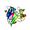
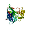
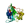
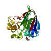
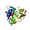
| Title | Structure of thaumatin collected by rotation serial crystallography on a COC membrane at a synchrotron source | ||||||
 Components Components | Thaumatin-1 | ||||||
 Keywords Keywords | PLANT PROTEIN | ||||||
| Function / homology |  Function and homology information Function and homology information | ||||||
| Biological species |  Thaumatococcus daniellii (katemfe) Thaumatococcus daniellii (katemfe) | ||||||
| Method |  X-RAY DIFFRACTION / SYNCHROTRON / MOLECULAR REPLACEMENT / Resolution: 1.65 Å X-RAY DIFFRACTION / SYNCHROTRON / MOLECULAR REPLACEMENT / Resolution: 1.65 Å | ||||||
 Authors Authors | Martiel, I. / Padeste, C. / Karpik, A. / Huang, C.Y. / Vera, L. / Wang, M. / Marsh, M. | ||||||
 Citation Citation | Journal: Acta Crystallogr D Struct Biol / Year: 2021 Title: Versatile microporous polymer-based supports for serial macromolecular crystallography. Authors: Martiel, I. / Beale, J.H. / Karpik, A. / Huang, C.Y. / Vera, L. / Olieric, N. / Wranik, M. / Tsai, C.J. / Muhle, J. / Aurelius, O. / John, J. / Hogbom, M. / Wang, M. / Marsh, M. / Padeste, C. | ||||||
| History |
|
- Structure visualization
| Structure viewer | Molecule: MolmilJmol/JSmol |
|---|
- Downloads & links
-Download
| PDBx/mmCIF format | 7ac3.cif.gz | 107 KB | Display | PDBx/mmCIF format |
|---|---|---|---|---|
| PDB format | pdb7ac3.ent.gz | 80.3 KB | Display | PDB format |
| PDBx/mmJSON format | 7ac3.json.gz | Tree view | PDBx/mmJSON format | |
| Others |  Other downloads Other downloads |
-Validation report
| Arichive directory | https://data.pdbj.org/pub/pdb/validation_reports/ac/7ac3ftp://data.pdbj.org/pub/pdb/validation_reports/ac/7ac3 | HTTPS FTP |
|---|
-Related structure data
| Related structure data |  7ac2C  7ac4C  7ac5C  7ac6C  7ai8C  7ai9C  4axrS S: Starting model for refinement C: citing same article ( |
|---|---|
| Similar structure data |


-Links
 PDBj
PDBj

- Assembly
| Deposited unit | 
| ||||||||
|---|---|---|---|---|---|---|---|---|---|
| 1 |
| ||||||||
| Unit cell |
|
-Components
| #1: Protein | Mass: 22227.059 Da / Num. of mol.: 1 Source method: isolated from a genetically manipulated source Source: (gene. exp.) Thaumatococcus daniellii (katemfe) / Production host:  | ||||||||
|---|---|---|---|---|---|---|---|---|---|
| #2: Chemical | ChemComp-TLA /   Mass: 150.087 Da / Num. of mol.: 1 / Source method: obtained synthetically / Formula: C4H6O6 Mass: 150.087 Da / Num. of mol.: 1 / Source method: obtained synthetically / Formula: C4H6O6 | ||||||||
| #3: Chemical |   Mass: 76.094 Da / Num. of mol.: 2 / Source method: obtained synthetically / Formula: C3H8O2 Mass: 76.094 Da / Num. of mol.: 2 / Source method: obtained synthetically / Formula: C3H8O2#4: Chemical |   Mass: 22.990 Da / Num. of mol.: 2 / Source method: obtained synthetically / Formula: Na Mass: 22.990 Da / Num. of mol.: 2 / Source method: obtained synthetically / Formula: Na#5: Water | ChemComp-HOH / |  Mass: 18.015 Da / Num. of mol.: 291 / Source method: isolated from a natural source / Formula: H2O Mass: 18.015 Da / Num. of mol.: 291 / Source method: isolated from a natural source / Formula: H2OHas ligand of interest | N | Has protein modification | Y | |
-Experimental details
-Experiment
| Experiment | Method: X-RAY DIFFRACTION / Number of used crystals: 1 |
|---|
- Sample preparation
| Crystal | Density Matthews: 2.84 Å3/Da / Density % sol: 56.75 % |
|---|---|
| Crystal grow | Temperature: 293 K / Method: batch mode Details: 1.6 M potassium sodium tartrate tetrahydrate, 100mM Na Hepes pH 7.0 |
-Data collection
| Diffraction | Mean temperature: 100 K / Serial crystal experiment: N |
|---|---|
| Diffraction source | Source: SYNCHROTRON / Site: SLS  / Beamline: X06SA / Wavelength: 1 Å / Beamline: X06SA / Wavelength: 1 Å |
| Detector | Type: DECTRIS EIGER X 16M / Detector: PIXEL / Date: Oct 10, 2018 |
| Radiation | Protocol: SINGLE WAVELENGTH / Monochromatic (M) / Laue (L): M / Scattering type: x-ray |
| Radiation wavelength | Wavelength: 1 Å / Relative weight: 1 |
| Reflection | Resolution: 1.65→45.92 Å / Num. obs: 31814 / % possible obs: 99.5 % / Redundancy: 7.98 % / CC1/2: 0.986 / Net I/σ(I): 4.19 |
| Reflection shell | Resolution: 1.65→1.69 Å / Mean I/σ(I) obs: 0.54 / Num. unique obs: 3127 / CC1/2: 0.144 |
- Processing
| Software |
| ||||||||||||||||||||||||||||||||||||||||||||||||||||||||||||
|---|---|---|---|---|---|---|---|---|---|---|---|---|---|---|---|---|---|---|---|---|---|---|---|---|---|---|---|---|---|---|---|---|---|---|---|---|---|---|---|---|---|---|---|---|---|---|---|---|---|---|---|---|---|---|---|---|---|---|---|---|---|
| Refinement | Method to determine structure: MOLECULAR REPLACEMENT Starting model: 4AXR Resolution: 1.65→45.916 Å / SU ML: 0.24 / Cross valid method: THROUGHOUT / σ(F): 1.33 / Phase error: 21.06 / Stereochemistry target values: ML
| ||||||||||||||||||||||||||||||||||||||||||||||||||||||||||||
| Solvent computation | Shrinkage radii: 0.9 Å / VDW probe radii: 1.11 Å / Solvent model: FLAT BULK SOLVENT MODEL | ||||||||||||||||||||||||||||||||||||||||||||||||||||||||||||
| Displacement parameters | Biso max: 66.35 Å2 / Biso mean: 26.2115 Å2 / Biso min: 14.42 Å2 | ||||||||||||||||||||||||||||||||||||||||||||||||||||||||||||
| Refinement step | Cycle: final / Resolution: 1.65→45.916 Å
| ||||||||||||||||||||||||||||||||||||||||||||||||||||||||||||
| LS refinement shell | Refine-ID: X-RAY DIFFRACTION / Rfactor Rfree error: 0 / % reflection obs: 100 %
| ||||||||||||||||||||||||||||||||||||||||||||||||||||||||||||
| Refinement TLS params. | Method: refined / Origin x: 4.6149 Å / Origin y: -16.6534 Å / Origin z: -4.8596 Å
| ||||||||||||||||||||||||||||||||||||||||||||||||||||||||||||
| Refinement TLS group |
|
