 Movie
Movie Controller

[English] 日本語
 Yorodumi
- PDB-7ai8: Structure of Ribonucleotide reductase R2 from Escherichia coli co... -
+ Open data

- Basic information
| Entry | Database: PDB / ID: 7ai8 | |||||||||
|---|---|---|---|---|---|---|---|---|---|---|
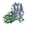
| Title | Structure of Ribonucleotide reductase R2 from Escherichia coli collected by still serial crystallography on a COC membrane at a synchrotron source | |||||||||
 Components Components | Ribonucleoside-diphosphate reductase 1 subunit beta | |||||||||
 Keywords Keywords | OXIDOREDUCTASE / metalloprotein | |||||||||
| Function / homology |  Function and homology information Function and homology informationribonucleoside diphosphate metabolic process / 2'-deoxyribonucleotide biosynthetic process / nucleobase-containing small molecule interconversion / ribonucleoside-diphosphate reductase complex / ribonucleoside-diphosphate reductase / ribonucleoside-diphosphate reductase activity, thioredoxin disulfide as acceptor / deoxyribonucleotide biosynthetic process / iron ion binding / identical protein binding / cytoplasm / cytosol Similarity search - Function | |||||||||
| Biological species |  | |||||||||
| Method |  X-RAY DIFFRACTION / SYNCHROTRON / MOLECULAR REPLACEMENT / Resolution: 2.1 Å X-RAY DIFFRACTION / SYNCHROTRON / MOLECULAR REPLACEMENT / Resolution: 2.1 Å | |||||||||
 Authors Authors | Aurelius, O. / John, J. / Martiel, I. / Padeste, C. / Karpik, A. / Huang, C.Y. / Hogbom, M. / Wang, M. / Marsh, M. | |||||||||
| Funding support |  Sweden, European Union, 2items Sweden, European Union, 2items
| |||||||||
 Citation Citation | Journal: Acta Crystallogr D Struct Biol / Year: 2021 Title: Versatile microporous polymer-based supports for serial macromolecular crystallography. Authors: Martiel, I. / Beale, J.H. / Karpik, A. / Huang, C.Y. / Vera, L. / Olieric, N. / Wranik, M. / Tsai, C.J. / Muhle, J. / Aurelius, O. / John, J. / Hogbom, M. / Wang, M. / Marsh, M. / Padeste, C. | |||||||||
| History |
|
- Structure visualization
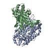
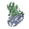
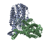
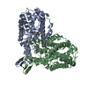
| Structure viewer | Molecule: MolmilJmol/JSmol |
|---|
- Downloads & links
-Download
| PDBx/mmCIF format | 7ai8.cif.gz | 175.2 KB | Display | PDBx/mmCIF format |
|---|---|---|---|---|
| PDB format | pdb7ai8.ent.gz | 123.1 KB | Display | PDB format |
| PDBx/mmJSON format | 7ai8.json.gz | Tree view | PDBx/mmJSON format | |
| Others |  Other downloads Other downloads |
-Validation report
| Arichive directory | https://data.pdbj.org/pub/pdb/validation_reports/ai/7ai8ftp://data.pdbj.org/pub/pdb/validation_reports/ai/7ai8 | HTTPS FTP |
|---|
-Related structure data
| Related structure data |  7ac2C  7ac3C 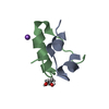 7ac4C 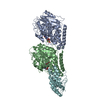 7ac5C 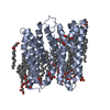 7ac6C  7ai9C  1mxrS C: citing same article ( S: Starting model for refinement |
|---|---|
| Similar structure data |


-Links
 PDBj
PDBj
- Assembly
| Deposited unit | 
| ||||||||||||
|---|---|---|---|---|---|---|---|---|---|---|---|---|---|
| 1 | 
| ||||||||||||
| Unit cell |
| ||||||||||||
| Components on special symmetry positions |
|
-Components
| #1: Protein | Mass: 43558.055 Da / Num. of mol.: 1 Source method: isolated from a genetically manipulated source Source: (gene. exp.) Strain: K12 / Gene: nrdB, ftsB, b2235, JW2229 / Production host: References: UniProt: P69924, ribonucleoside-diphosphate reductase | ||||
|---|---|---|---|---|---|
| #2: Chemical |   Mass: 55.845 Da / Num. of mol.: 2 / Source method: obtained synthetically / Formula: Fe / Feature type: SUBJECT OF INVESTIGATION Mass: 55.845 Da / Num. of mol.: 2 / Source method: obtained synthetically / Formula: Fe / Feature type: SUBJECT OF INVESTIGATION#3: Water | ChemComp-HOH / |  Mass: 18.015 Da / Num. of mol.: 103 / Source method: isolated from a natural source / Formula: H2O Mass: 18.015 Da / Num. of mol.: 103 / Source method: isolated from a natural source / Formula: H2OHas ligand of interest | Y | |
-Experimental details
-Experiment
| Experiment | Method: X-RAY DIFFRACTION / Number of used crystals: 1 |
|---|
- Sample preparation
| Crystal | Density Matthews: 2.82 Å3/Da / Density % sol: 56.39 % |
|---|---|
| Crystal grow | Temperature: 294 K / Method: vapor diffusion, sitting drop / pH: 7 Details: 34-37 % PAA 2100, 100 mM HEPES 7.0, 250-450 mM NaCl, 200 mM ammonium sulfate [and] 26% PAA 2100, 100 mM HEPES 7.0, 150 NaCl, 100 Malonate |
-Data collection
| Diffraction | Mean temperature: 100 K / Serial crystal experiment: Y |
|---|---|
| Diffraction source | Source: SYNCHROTRON / Site: SLS  / Beamline: X06SA / Wavelength: 1 Å / Beamline: X06SA / Wavelength: 1 Å |
| Detector | Type: DECTRIS EIGER X 16M / Detector: PIXEL / Date: Mar 14, 2019 |
| Radiation | Protocol: SINGLE WAVELENGTH / Monochromatic (M) / Laue (L): M / Scattering type: x-ray |
| Radiation wavelength | Wavelength: 1 Å / Relative weight: 1 |
| Reflection | Resolution: 2.08→52.2 Å / Num. obs: 31083 / % possible obs: 99.65 % / Redundancy: 201.59 % / Biso Wilson estimate: 41.87 Å2 / CC1/2: 0.9967 / CC star: 0.9992 / R split: 0.0538 / Net I/σ(I): 10.74 |
| Reflection shell | Resolution: 2.1→2.13 Å / Redundancy: 15.2 % / Mean I/σ(I) obs: 1.74 / Num. unique obs: 1474 / CC1/2: 0.3829 / CC star: 0.7441 / R split: 0.7483 / % possible all: 96.66 |
| Serial crystallography sample delivery | Method: fixed target |
| Serial crystallography sample delivery fixed target | Sample holding: COC thin membrane |
- Processing
| Software |
| ||||||||||||||||||||||||||||||||||||||||||||||||||||||||||||||||||||||||||||||||||||
|---|---|---|---|---|---|---|---|---|---|---|---|---|---|---|---|---|---|---|---|---|---|---|---|---|---|---|---|---|---|---|---|---|---|---|---|---|---|---|---|---|---|---|---|---|---|---|---|---|---|---|---|---|---|---|---|---|---|---|---|---|---|---|---|---|---|---|---|---|---|---|---|---|---|---|---|---|---|---|---|---|---|---|---|---|---|
| Refinement | Method to determine structure: MOLECULAR REPLACEMENT Starting model: 1MXR Resolution: 2.1→51.97 Å / SU ML: 0.2272 / Cross valid method: FREE R-VALUE / σ(F): 1.33 / Phase error: 24.9367 Stereochemistry target values: GeoStd + Monomer Library + CDL v1.2
| ||||||||||||||||||||||||||||||||||||||||||||||||||||||||||||||||||||||||||||||||||||
| Solvent computation | Shrinkage radii: 0.9 Å / VDW probe radii: 1.11 Å / Solvent model: FLAT BULK SOLVENT MODEL | ||||||||||||||||||||||||||||||||||||||||||||||||||||||||||||||||||||||||||||||||||||
| Displacement parameters | Biso mean: 57.43 Å2 | ||||||||||||||||||||||||||||||||||||||||||||||||||||||||||||||||||||||||||||||||||||
| Refinement step | Cycle: LAST / Resolution: 2.1→51.97 Å
| ||||||||||||||||||||||||||||||||||||||||||||||||||||||||||||||||||||||||||||||||||||
| Refine LS restraints |
| ||||||||||||||||||||||||||||||||||||||||||||||||||||||||||||||||||||||||||||||||||||
| LS refinement shell |
| ||||||||||||||||||||||||||||||||||||||||||||||||||||||||||||||||||||||||||||||||||||
| Refinement TLS params. | Method: refined / Origin x: -21.6870560366 Å / Origin y: 29.9104760991 Å / Origin z: 4.74723600776 Å
| ||||||||||||||||||||||||||||||||||||||||||||||||||||||||||||||||||||||||||||||||||||
| Refinement TLS group | Selection details: (chain 'A' and resid 1 through 339) |
