 Movie
Movie Controller


+ Open data

- Basic information
| Entry | Database: PDB / ID: 7aah | ||||||
|---|---|---|---|---|---|---|---|
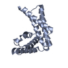
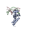
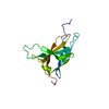
| Title | Structure of human pERp1 | ||||||
 Components Components | Marginal zone B- and B1-cell-specific protein | ||||||
 Keywords Keywords | IMMUNE SYSTEM / Folding factor / Endoplasmatic Reticulum / IgM | ||||||
| Function / homology |  Function and homology information Function and homology informationregulation of B cell proliferation / regulation of insulin receptor signaling pathway / endoplasmic reticulum chaperone complex / integrin activation / positive regulation of immunoglobulin production / regulation of cell population proliferation / endoplasmic reticulum lumen / positive regulation of cell population proliferation / apoptotic process / extracellular region / cytoplasm Similarity search - Function | ||||||
| Biological species |  Homo sapiens (human) Homo sapiens (human) | ||||||
| Method |  X-RAY DIFFRACTION / SYNCHROTRON / AB INITIO PHASING / Resolution: 1.4 Å X-RAY DIFFRACTION / SYNCHROTRON / AB INITIO PHASING / Resolution: 1.4 Å | ||||||
 Authors Authors | Sowa, S.T. / Moilanen, A. / Biterova, E. / Saaranen, M.J. / Lehtio, L. / Ruddock, L.W. | ||||||
| Funding support |  Finland, 1items Finland, 1items
| ||||||
 Citation Citation | Journal: J.Mol.Biol. / Year: 2021 Title: High-resolution Crystal Structure of Human pERp1, A Saposin-like Protein Involved in IgA, IgM and Integrin Maturation in the Endoplasmic Reticulum. Authors: Sowa, S.T. / Moilanen, A. / Biterova, E. / Saaranen, M.J. / Lehtio, L. / Ruddock, L.W. | ||||||
| History |
|
- Structure visualization
| Structure viewer | Molecule: MolmilJmol/JSmol |
|---|
- Downloads & links
-Download
| PDBx/mmCIF format | 7aah.cif.gz | 192.8 KB | Display | PDBx/mmCIF format |
|---|---|---|---|---|
| PDB format | pdb7aah.ent.gz | 156 KB | Display | PDB format |
| PDBx/mmJSON format | 7aah.json.gz | Tree view | PDBx/mmJSON format | |
| Others |  Other downloads Other downloads |
-Validation report
| Arichive directory | https://data.pdbj.org/pub/pdb/validation_reports/aa/7aahftp://data.pdbj.org/pub/pdb/validation_reports/aa/7aah | HTTPS FTP |
|---|
-Related structure data
| Similar structure data |
|---|


-Links
 PDBj
PDBj

- Assembly
| Deposited unit | 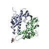
| ||||||||
|---|---|---|---|---|---|---|---|---|---|
| 1 | 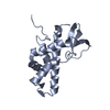
| ||||||||
| 2 | 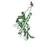
| ||||||||
| Unit cell |
|
-Components
| #1: Protein | Mass: 18257.389 Da / Num. of mol.: 2 Source method: isolated from a genetically manipulated source Source: (gene. exp.) Homo sapiens (human) / Gene: MZB1, MEDA7, PACAP, HSPC190 / Production host:  #2: Chemical | ChemComp-CIT / |   Mass: 192.124 Da / Num. of mol.: 1 / Source method: obtained synthetically / Formula: C6H8O7 Mass: 192.124 Da / Num. of mol.: 1 / Source method: obtained synthetically / Formula: C6H8O7#3: Water | ChemComp-HOH / |  Mass: 18.015 Da / Num. of mol.: 312 / Source method: isolated from a natural source / Formula: H2O Mass: 18.015 Da / Num. of mol.: 312 / Source method: isolated from a natural source / Formula: H2OHas ligand of interest | N | Has protein modification | Y | |
|---|
-Experimental details
-Experiment
| Experiment | Method: X-RAY DIFFRACTION / Number of used crystals: 1 |
|---|
- Sample preparation
| Crystal | Density Matthews: 2 Å3/Da / Density % sol: 38.63 % |
|---|---|
| Crystal grow | Temperature: 293.15 K / Method: vapor diffusion, sitting drop Details: Lithium citrate tribasic tetrahydrate, Glycerol ethoxylate, n-decyl-beta-D-maltopyranoside |
-Data collection
| Diffraction | Mean temperature: 100 K / Serial crystal experiment: N |
|---|---|
| Diffraction source | Source: SYNCHROTRON / Site: Diamond  / Beamline: I24 / Wavelength: 0.96863 Å / Beamline: I24 / Wavelength: 0.96863 Å |
| Detector | Type: DECTRIS PILATUS 6M / Detector: PIXEL / Date: Oct 6, 2017 |
| Radiation | Protocol: SINGLE WAVELENGTH / Monochromatic (M) / Laue (L): M / Scattering type: x-ray |
| Radiation wavelength | Wavelength: 0.96863 Å / Relative weight: 1 |
| Reflection | Resolution: 1.4→47.8 Å / Num. obs: 58099 / % possible obs: 99.1 % / Redundancy: 6.2 % / CC1/2: 0.999 / Net I/σ(I): 12.88 |
| Reflection shell | Resolution: 1.4→1.45 Å / Num. unique obs: 5426 / CC1/2: 0.364 |
- Processing
| Software |
| |||||||||||||||||||||||||||||||||||||||||||||||||||||||||||||||||||||||||||||||||||||||||||||||||||||||||||||||||||||||||||||||||||||||||||||||||||
|---|---|---|---|---|---|---|---|---|---|---|---|---|---|---|---|---|---|---|---|---|---|---|---|---|---|---|---|---|---|---|---|---|---|---|---|---|---|---|---|---|---|---|---|---|---|---|---|---|---|---|---|---|---|---|---|---|---|---|---|---|---|---|---|---|---|---|---|---|---|---|---|---|---|---|---|---|---|---|---|---|---|---|---|---|---|---|---|---|---|---|---|---|---|---|---|---|---|---|---|---|---|---|---|---|---|---|---|---|---|---|---|---|---|---|---|---|---|---|---|---|---|---|---|---|---|---|---|---|---|---|---|---|---|---|---|---|---|---|---|---|---|---|---|---|---|---|---|---|
| Refinement | Method to determine structure: AB INITIO PHASING / Resolution: 1.4→47.764 Å / SU ML: 0.18 / Cross valid method: FREE R-VALUE / σ(F): 1.33 / Phase error: 20.32 / Stereochemistry target values: ML
| |||||||||||||||||||||||||||||||||||||||||||||||||||||||||||||||||||||||||||||||||||||||||||||||||||||||||||||||||||||||||||||||||||||||||||||||||||
| Solvent computation | Shrinkage radii: 0.9 Å / VDW probe radii: 1.11 Å / Solvent model: FLAT BULK SOLVENT MODEL | |||||||||||||||||||||||||||||||||||||||||||||||||||||||||||||||||||||||||||||||||||||||||||||||||||||||||||||||||||||||||||||||||||||||||||||||||||
| Refinement step | Cycle: LAST / Resolution: 1.4→47.764 Å
| |||||||||||||||||||||||||||||||||||||||||||||||||||||||||||||||||||||||||||||||||||||||||||||||||||||||||||||||||||||||||||||||||||||||||||||||||||
| Refine LS restraints |
| |||||||||||||||||||||||||||||||||||||||||||||||||||||||||||||||||||||||||||||||||||||||||||||||||||||||||||||||||||||||||||||||||||||||||||||||||||
| LS refinement shell |
|
