 Movie
Movie Controller


+ Open data

- Basic information
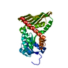
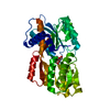
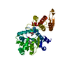
| Entry | Database: PDB / ID: 7a90 | ||||||
|---|---|---|---|---|---|---|---|
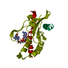
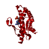
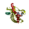
| Title | WT STING in complex with 3',3'-c-di[2'FdAM(PS)] | ||||||
 Components Components | Stimulator of interferon protein | ||||||
 Keywords Keywords | PROTEIN BINDING / Innate immune system / cyclic dinucleotides | ||||||
| Function / homology |  Function and homology information Function and homology information2',3'-cyclic GMP-AMP binding / cyclic-di-GMP binding / reticulophagy / positive regulation of type I interferon production / autophagosome membrane / positive regulation of macroautophagy / autophagosome assembly / activation of innate immune response / endoplasmic reticulum-Golgi intermediate compartment membrane / monoatomic ion transmembrane transport ...2',3'-cyclic GMP-AMP binding / cyclic-di-GMP binding / reticulophagy / positive regulation of type I interferon production / autophagosome membrane / positive regulation of macroautophagy / autophagosome assembly / activation of innate immune response / endoplasmic reticulum-Golgi intermediate compartment membrane / monoatomic ion transmembrane transport / cytoplasmic vesicle / defense response to virus / mitochondrial outer membrane / Golgi membrane / innate immune response / endoplasmic reticulum membrane / perinuclear region of cytoplasm Similarity search - Function | ||||||
| Biological species |  Homo sapiens (human) Homo sapiens (human) | ||||||
| Method |  X-RAY DIFFRACTION / MOLECULAR REPLACEMENT / molecular replacement / Resolution: 3.185 Å X-RAY DIFFRACTION / MOLECULAR REPLACEMENT / molecular replacement / Resolution: 3.185 Å | ||||||
 Authors Authors | Boura, E. / Smola, M. | ||||||
 Citation Citation | Journal: To Be Published Title: WT STING in complex with 3',3'-c-di[2'FdAM(PS)] Authors: Boura, E. / Smola, M. | ||||||
| History |
|
- Structure visualization
| Structure viewer | Molecule: MolmilJmol/JSmol |
|---|
- Downloads & links
-Download
| PDBx/mmCIF format | 7a90.cif.gz | 77.5 KB | Display | PDBx/mmCIF format |
|---|---|---|---|---|
| PDB format | pdb7a90.ent.gz | 57.2 KB | Display | PDB format |
| PDBx/mmJSON format | 7a90.json.gz | Tree view | PDBx/mmJSON format | |
| Others |  Other downloads Other downloads |
-Validation report
| Summary document | 7a90_validation.pdf.gz | 827.7 KB | Display | wwPDB validaton report |
|---|---|---|---|---|
| Full document | 7a90_full_validation.pdf.gz | 830.1 KB | Display | |
| Data in XML | 7a90_validation.xml.gz | 6.3 KB | Display | |
| Data in CIF | 7a90_validation.cif.gz | 8.1 KB | Display | |
| Arichive directory | https://data.pdbj.org/pub/pdb/validation_reports/a9/7a90ftp://data.pdbj.org/pub/pdb/validation_reports/a9/7a90 | HTTPS FTP |
-Related structure data
| Related structure data |  4ksyS S: Starting model for refinement |
|---|---|
| Similar structure data |

-Links
 PDBj
PDBj- Assembly
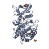
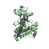
| Deposited unit | 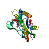
| ||||||||
|---|---|---|---|---|---|---|---|---|---|
| 1 | 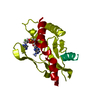
| ||||||||
| Unit cell |
|
-Components
| #1: Protein | Mass: 23189.064 Da / Num. of mol.: 1 Source method: isolated from a genetically manipulated source Source: (gene. exp.) Homo sapiens (human) / Gene: STING, LOC340061, hCG_1782396 / Production host:  |
|---|---|
| #2: Chemical | ChemComp-R4T / 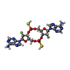  Mass: 696.541 Da / Num. of mol.: 1 / Source method: obtained synthetically / Formula: C20H24F2N10O8P2S2 / Feature type: SUBJECT OF INVESTIGATION Mass: 696.541 Da / Num. of mol.: 1 / Source method: obtained synthetically / Formula: C20H24F2N10O8P2S2 / Feature type: SUBJECT OF INVESTIGATION |
| Has ligand of interest | Y |
-Experimental details
-Experiment
| Experiment | Method: X-RAY DIFFRACTION / Number of used crystals: 1 |
|---|
- Sample preparation
| Crystal | Density Matthews: 2.36 Å3/Da / Density % sol: 47.8 % |
|---|---|
| Crystal grow | Temperature: 291 K / Method: vapor diffusion, sitting drop Details: 0.2 M tri-Potassium Citrate monohydrate, 20% (w/v) PEG 3350, 0.1 M Natrium Citrate, 50 mM EDTA |
-Data collection
| Diffraction | Mean temperature: 100 K / Serial crystal experiment: N |
|---|---|
| Diffraction source | Source: ROTATING ANODE / Type: RIGAKU MICROMAX-007 HF / Wavelength: 1.54 Å |
| Detector | Type: DECTRIS PILATUS 200K / Detector: PIXEL / Date: Jul 27, 2020 |
| Radiation | Protocol: SINGLE WAVELENGTH / Monochromatic (M) / Laue (L): M / Scattering type: x-ray |
| Radiation wavelength | Wavelength: 1.54 Å / Relative weight: 1 |
| Reflection | Resolution: 3.185→35.042 Å / Num. obs: 4032 / % possible obs: 99.8 % / Redundancy: 11.6 % / CC1/2: 0.967 / Net I/σ(I): 5.84 |
| Reflection shell | Resolution: 3.185→3.299 Å / Mean I/σ(I) obs: 1.91 / Num. unique obs: 390 / CC1/2: 0.678 |
-Phasing
| Phasing | Method: molecular replacement | |||||||||
|---|---|---|---|---|---|---|---|---|---|---|
| Phasing MR |
|
- Processing
| Software |
| ||||||||||||||||||||||||
|---|---|---|---|---|---|---|---|---|---|---|---|---|---|---|---|---|---|---|---|---|---|---|---|---|---|
| Refinement | Method to determine structure: MOLECULAR REPLACEMENT Starting model: 4KSY Resolution: 3.185→35.04 Å / SU ML: 0.4 / Cross valid method: THROUGHOUT / σ(F): 1.34 / Phase error: 16.31 / Stereochemistry target values: ML
| ||||||||||||||||||||||||
| Solvent computation | Shrinkage radii: 0.9 Å / VDW probe radii: 1.11 Å / Solvent model: FLAT BULK SOLVENT MODEL | ||||||||||||||||||||||||
| Displacement parameters | Biso max: 95.81 Å2 / Biso mean: 33.5966 Å2 / Biso min: 13.81 Å2 | ||||||||||||||||||||||||
| Refinement step | Cycle: final / Resolution: 3.185→35.04 Å
| ||||||||||||||||||||||||
| LS refinement shell | Resolution: 3.185→3.299 Å / Rfactor Rfree error: 0
|
