 Movie
Movie Controller

[English] 日本語
 Yorodumi
- PDB-1jx6: CRYSTAL STRUCTURE OF LUXP FROM VIBRIO HARVEYI COMPLEXED WITH AUTO... -
+ Open data

- Basic information
| Entry | Database: PDB / ID: 1jx6 | ||||||
|---|---|---|---|---|---|---|---|
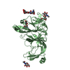
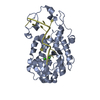
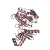
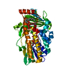
| Title | CRYSTAL STRUCTURE OF LUXP FROM VIBRIO HARVEYI COMPLEXED WITH AUTOINDUCER-2 | ||||||
 Components Components | LUXP PROTEIN | ||||||
 Keywords Keywords | SIGNALING PROTEIN / PROTEIN-LIGAND COMPLEX | ||||||
| Function / homology |  Function and homology information Function and homology informationtransmembrane transport / periplasmic space / transcription cis-regulatory region binding / DNA-binding transcription factor activity Similarity search - Function | ||||||
| Biological species |  Vibrio harveyi (bacteria) Vibrio harveyi (bacteria) | ||||||
| Method |  X-RAY DIFFRACTION / SYNCHROTRON / MIR / Resolution: 1.5 Å X-RAY DIFFRACTION / SYNCHROTRON / MIR / Resolution: 1.5 Å | ||||||
 Authors Authors | Chen, X. / Schauder, S. / Potier, N. / Van Dorsselaer, A. / Pelczer, I. / BassleR, B.L. / Hughson, F.M. | ||||||
 Citation Citation | Journal: Nature / Year: 2002 Title: Structural identification of a bacterial quorum-sensing signal containing boron. Authors: Chen, X. / Schauder, S. / Potier, N. / Van Dorsselaer, A. / Pelczer, I. / Bassler, B.L. / Hughson, F.M. | ||||||
| History |
|
- Structure visualization
| Structure viewer | Molecule: MolmilJmol/JSmol |
|---|
- Downloads & links
-Download
| PDBx/mmCIF format | 1jx6.cif.gz | 88.9 KB | Display | PDBx/mmCIF format |
|---|---|---|---|---|
| PDB format | pdb1jx6.ent.gz | 65.8 KB | Display | PDB format |
| PDBx/mmJSON format | 1jx6.json.gz | Tree view | PDBx/mmJSON format | |
| Others |  Other downloads Other downloads |
-Validation report
| Arichive directory | https://data.pdbj.org/pub/pdb/validation_reports/jx/1jx6ftp://data.pdbj.org/pub/pdb/validation_reports/jx/1jx6 | HTTPS FTP |
|---|
-Related structure data
| Similar structure data |
|---|


-Links
 PDBj
PDBj- Assembly
| Deposited unit | 
| ||||||||||
|---|---|---|---|---|---|---|---|---|---|---|---|
| 1 |
| ||||||||||
| Unit cell |
| ||||||||||
| Details | PROBABLY MONOMER |
-Components
| #1: Protein | Mass: 39065.734 Da / Num. of mol.: 1 Source method: isolated from a genetically manipulated source Source: (gene. exp.) Vibrio harveyi (bacteria) / Gene: LUXP / Plasmid: PGEX4T / Species (production host): Escherichia coli / Production host: |
|---|---|
| #2: Chemical | ChemComp-CA /   Mass: 40.078 Da / Num. of mol.: 1 / Source method: obtained synthetically / Formula: Ca Mass: 40.078 Da / Num. of mol.: 1 / Source method: obtained synthetically / Formula: Ca |
| #3: Chemical | ChemComp-AI2 /   Mass: 192.940 Da / Num. of mol.: 1 / Source method: obtained synthetically / Formula: C5H10BO7 Mass: 192.940 Da / Num. of mol.: 1 / Source method: obtained synthetically / Formula: C5H10BO7 |
| #4: Water | ChemComp-HOH /  Mass: 18.015 Da / Num. of mol.: 332 / Source method: isolated from a natural source / Formula: H2O Mass: 18.015 Da / Num. of mol.: 332 / Source method: isolated from a natural source / Formula: H2O |
-Experimental details
-Experiment
| Experiment | Method: X-RAY DIFFRACTION / Number of used crystals: 1 |
|---|
- Sample preparation
| Crystal | Density Matthews: 2.27 Å3/Da / Density % sol: 45.76 % | ||||||||||||||||||||||||||||||
|---|---|---|---|---|---|---|---|---|---|---|---|---|---|---|---|---|---|---|---|---|---|---|---|---|---|---|---|---|---|---|---|
| Crystal grow | Temperature: 298 K / Method: vapor diffusion, hanging drop / pH: 8.5 Details: PEG 4000, GLYCEROL, pH 8.5, VAPOR DIFFUSION, HANGING DROP at 298K | ||||||||||||||||||||||||||||||
| Crystal grow | *PLUS Method: unknown | ||||||||||||||||||||||||||||||
| Components of the solutions | *PLUS
|
-Data collection
| Diffraction | Mean temperature: 100 K |
|---|---|
| Diffraction source | Source: SYNCHROTRON / Site: NSLS  / Beamline: X12C / Wavelength: 1.072 Å / Beamline: X12C / Wavelength: 1.072 Å |
| Detector | Type: BRANDEIS / Detector: CCD / Date: Apr 14, 2001 |
| Radiation | Monochromator: GRAPHITE / Protocol: SINGLE WAVELENGTH / Monochromatic (M) / Laue (L): M / Scattering type: x-ray |
| Radiation wavelength | Wavelength: 1.072 Å / Relative weight: 1 |
| Reflection | Resolution: 1.45→25 Å / Num. all: 61620 / Num. obs: 61620 / % possible obs: 100 % / Observed criterion σ(F): 0 / Observed criterion σ(I): 0 / Biso Wilson estimate: 20.3 Å2 / Rmerge(I) obs: 0.04 / Rsym value: 0.036 / Net I/σ(I): 42 |
| Reflection shell | Resolution: 1.45→1.5 Å / Rmerge(I) obs: 0.297 / Mean I/σ(I) obs: 3.6 / Num. unique all: 6131 / Rsym value: 0.297 / % possible all: 100 |
| Reflection | *PLUS % possible obs: 100 % / Redundancy: 5.5 % / Rmerge(I) obs: 0.035 |
| Reflection shell | *PLUS Rmerge(I) obs: 0.25 |
- Processing
| Software |
| ||||||||||||||||||||||||||||||||||||||||
|---|---|---|---|---|---|---|---|---|---|---|---|---|---|---|---|---|---|---|---|---|---|---|---|---|---|---|---|---|---|---|---|---|---|---|---|---|---|---|---|---|---|
| Refinement | Method to determine structure: MIR / Resolution: 1.5→24.58 Å / Rfactor Rfree error: 0.004 / Data cutoff high absF: 542802.82 / Data cutoff low absF: 0 / Isotropic thermal model: RESTRAINED / Cross valid method: THROUGHOUT / σ(F): 1 / Stereochemistry target values: Engh & Huber
| ||||||||||||||||||||||||||||||||||||||||
| Solvent computation | Solvent model: FLAT MODEL / Bsol: 55.4898 Å2 / ksol: 0.358532 e/Å3 | ||||||||||||||||||||||||||||||||||||||||
| Displacement parameters | Biso mean: 25.3 Å2
| ||||||||||||||||||||||||||||||||||||||||
| Refine analyze |
| ||||||||||||||||||||||||||||||||||||||||
| Refinement step | Cycle: LAST / Resolution: 1.5→24.58 Å
| ||||||||||||||||||||||||||||||||||||||||
| Refine LS restraints |
| ||||||||||||||||||||||||||||||||||||||||
| LS refinement shell | Resolution: 1.5→1.55 Å / Rfactor Rfree error: 0.017 / Total num. of bins used: 10
| ||||||||||||||||||||||||||||||||||||||||
| Xplor file |
| ||||||||||||||||||||||||||||||||||||||||
| Software | *PLUS Name: CNS / Version: 1 / Classification: refinement | ||||||||||||||||||||||||||||||||||||||||
| Refinement | *PLUS Lowest resolution: 25 Å / Num. reflection obs: 53816 / σ(F): 1 / % reflection Rfree: 8.1 % | ||||||||||||||||||||||||||||||||||||||||
| Solvent computation | *PLUS | ||||||||||||||||||||||||||||||||||||||||
| Displacement parameters | *PLUS Biso mean: 25.3 Å2 | ||||||||||||||||||||||||||||||||||||||||
| Refine LS restraints | *PLUS
| ||||||||||||||||||||||||||||||||||||||||
| LS refinement shell | *PLUS Rfactor Rfree: 0.317 / % reflection Rfree: 8.3 % / Rfactor Rwork: 0.274 / Rfactor obs: 0.274 |
