 Movie
Movie Controller


+ Open data

- Basic information
| Entry | Database: PDB / ID: 1gcu | ||||||
|---|---|---|---|---|---|---|---|
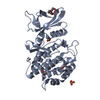
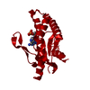
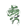
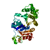
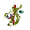
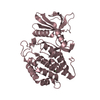
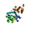
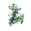
| Title | CRYSTAL STRUCTURE OF RAT BILIVERDIN REDUCTASE AT 1.4 A | ||||||
 Components Components | BILIVERDIN REDUCTASE A | ||||||
 Keywords Keywords | OXIDOREDUCTASE / biliverdin / Rossmann fold | ||||||
| Function / homology |  Function and homology information Function and homology informationbiliverdin reductase / biliberdin reductase (NADH) activity / biliverdin reductase (NADPH) activity / pigment metabolic process / biliverdin reductase [NAD(P)H] activity / Heme degradation / reactive oxygen species biosynthetic process / complement component C5a signaling pathway / Cytoprotection by HMOX1 / response to cyclosporin A ...biliverdin reductase / biliberdin reductase (NADH) activity / biliverdin reductase (NADPH) activity / pigment metabolic process / biliverdin reductase [NAD(P)H] activity / Heme degradation / reactive oxygen species biosynthetic process / complement component C5a signaling pathway / Cytoprotection by HMOX1 / response to cyclosporin A / lipid oxidation / sensory perception of itch / insulin metabolic process / apoptotic DNA fragmentation / ear development / heme catabolic process / triglyceride metabolic process / glycogen metabolic process / response to food / canonical NF-kappaB signal transduction / lipid metabolic process / phosphatidylinositol 3-kinase/protein kinase B signal transduction / fatty acid metabolic process / chemotaxis / insulin receptor signaling pathway / MAPK cascade / glucose homeostasis / response to oxidative stress / response to lipopolysaccharide / gene expression / immune response / RNA polymerase II cis-regulatory region sequence-specific DNA binding / inflammatory response / nucleotide binding / protein serine/threonine kinase activity / negative regulation of apoptotic process / cell surface / zinc ion binding / nucleus / cytoplasm / cytosol Similarity search - Function | ||||||
| Biological species |  | ||||||
| Method |  X-RAY DIFFRACTION / SYNCHROTRON / Resolution: 1.4 Å X-RAY DIFFRACTION / SYNCHROTRON / Resolution: 1.4 Å | ||||||
 Authors Authors | Kikuchi, A. / Park, S.Y. / Shiro, Y. | ||||||
 Citation Citation | Journal: Nat.Struct.Biol. / Year: 2001 Title: Crystal structure of rat biliverdin reductase. Authors: Kikuchi, A. / Park, S.Y. / Miyatake, H. / Sun, D. / Sato, M. / Yoshida, T. / Shiro, Y. | ||||||
| History |
|
- Structure visualization
| Structure viewer | Molecule: MolmilJmol/JSmol |
|---|
- Downloads & links
-Download
| PDBx/mmCIF format | 1gcu.cif.gz | 75.2 KB | Display | PDBx/mmCIF format |
|---|---|---|---|---|
| PDB format | pdb1gcu.ent.gz | 55.9 KB | Display | PDB format |
| PDBx/mmJSON format | 1gcu.json.gz | Tree view | PDBx/mmJSON format | |
| Others |  Other downloads Other downloads |
-Validation report
| Arichive directory | https://data.pdbj.org/pub/pdb/validation_reports/gc/1gcuftp://data.pdbj.org/pub/pdb/validation_reports/gc/1gcu | HTTPS FTP |
|---|
-Related structure data
| Similar structure data |
|---|


-Links
 PDBj
PDBj- Assembly
| Deposited unit | 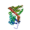
| ||||||||
|---|---|---|---|---|---|---|---|---|---|
| 1 |
| ||||||||
| Unit cell |
|
-Components
| #1: Protein | Mass: 33611.535 Da / Num. of mol.: 1 Source method: isolated from a genetically manipulated source Source: (gene. exp.)  |
|---|---|
| #2: Water | ChemComp-HOH /  Mass: 18.015 Da / Num. of mol.: 294 / Source method: isolated from a natural source / Formula: H2O Mass: 18.015 Da / Num. of mol.: 294 / Source method: isolated from a natural source / Formula: H2O |
-Experimental details
-Experiment
| Experiment | Method: X-RAY DIFFRACTION / Number of used crystals: 2 |
|---|
- Sample preparation
| Crystal | Density Matthews: 2.7 Å3/Da / Density % sol: 54.49 % | |||||||||||||||||||||||||
|---|---|---|---|---|---|---|---|---|---|---|---|---|---|---|---|---|---|---|---|---|---|---|---|---|---|---|
| Crystal grow | Temperature: 283 K / Method: vapor diffusion, sitting drop / pH: 6.5 Details: (NH4)2SO4, Na citrate, Na/K tartrate, pH 6.5, VAPOR DIFFUSION, SITTING DROP, temperature 283K | |||||||||||||||||||||||||
| Crystal | *PLUS Density % sol: 51 % | |||||||||||||||||||||||||
| Crystal grow | *PLUS | |||||||||||||||||||||||||
| Components of the solutions | *PLUS
|
-Data collection
| Diffraction | Mean temperature: 100 K |
|---|---|
| Diffraction source | Source: SYNCHROTRON / Site: SPring-8  / Beamline: BL44B2 / Wavelength: 0.7 / Beamline: BL44B2 / Wavelength: 0.7 |
| Detector | Type: MARRESEARCH / Detector: CCD / Date: Mar 20, 2000 |
| Radiation | Protocol: SINGLE WAVELENGTH / Monochromatic (M) / Laue (L): M / Scattering type: x-ray |
| Radiation wavelength | Wavelength: 0.7 Å / Relative weight: 1 |
| Reflection | Resolution: 1.4→32.71 Å / Num. all: 379020 / Num. obs: 379020 / Observed criterion σ(F): 0 / Redundancy: 5.3 % / Biso Wilson estimate: 18.3 Å2 / Rmerge(I) obs: 0.079 / Net I/σ(I): 6 |
| Reflection shell | Resolution: 1.4→1.48 Å / Redundancy: 3.4 % / Rmerge(I) obs: 0.324 / Num. unique all: 9812 / % possible all: 94.3 |
| Reflection | *PLUS Num. obs: 71182 / Num. measured all: 379020 |
- Processing
| Software |
| |||||||||||||||||||||||||
|---|---|---|---|---|---|---|---|---|---|---|---|---|---|---|---|---|---|---|---|---|---|---|---|---|---|---|
| Refinement | Resolution: 1.4→32.7 Å / σ(F): 0 / Stereochemistry target values: Engh & Huber
| |||||||||||||||||||||||||
| Refinement step | Cycle: LAST / Resolution: 1.4→32.7 Å
| |||||||||||||||||||||||||
| Refine LS restraints |
| |||||||||||||||||||||||||
| Software | *PLUS Name: X-PLOR / Version: 3.851 / Classification: refinement | |||||||||||||||||||||||||
| Refinement | *PLUS Highest resolution: 1.4 Å / Lowest resolution: 32.7 Å / σ(F): 0 | |||||||||||||||||||||||||
| Solvent computation | *PLUS | |||||||||||||||||||||||||
| Displacement parameters | *PLUS | |||||||||||||||||||||||||
| Refine LS restraints | *PLUS Type: x_angle_deg / Dev ideal: 1.6 |
