 Movie
Movie Controller


+ Open data

- Basic information
| Entry | Database: PDB / ID: 6zus | ||||||||||||||||||
|---|---|---|---|---|---|---|---|---|---|---|---|---|---|---|---|---|---|---|---|
| Title | Crystal structure of the effector Ecp11-1 from Fulvia fulva | ||||||||||||||||||
 Components Components | Extracellular protein 11-1 | ||||||||||||||||||
 Keywords Keywords | SIGNALING PROTEIN / AVIRULENCE PROTEIN / AVIRULENCE EFFECTOR PROTEIN / FUNGAL PROTEIN / Fulvia fulva | ||||||||||||||||||
| Function / homology | metal ion binding / DI(HYDROXYETHYL)ETHER / Ecp11-1 Function and homology information Function and homology information | ||||||||||||||||||
| Biological species |  Passalora fulva (fungus) Passalora fulva (fungus) | ||||||||||||||||||
| Method |  X-RAY DIFFRACTION / SYNCHROTRON / MOLECULAR REPLACEMENT / Resolution: 1.62 Å X-RAY DIFFRACTION / SYNCHROTRON / MOLECULAR REPLACEMENT / Resolution: 1.62 Å | ||||||||||||||||||
 Authors Authors | Lazar, N. / Mesarich, C. / Petit-Houdenot, Y. / Talbi, N. / Li de la Sierra-Gallay, I. / Zelie, E. / Blondeau, K. / Gracy, J. / Ollivier, B. / van de Wouw, A. ...Lazar, N. / Mesarich, C. / Petit-Houdenot, Y. / Talbi, N. / Li de la Sierra-Gallay, I. / Zelie, E. / Blondeau, K. / Gracy, J. / Ollivier, B. / van de Wouw, A. / Balesdent, M.H. / Idnurm, A. / van Tilbeurgh, H. / Fudal, I. | ||||||||||||||||||
| Funding support |  France, 5items France, 5items
| ||||||||||||||||||
 Citation Citation | Journal: Plos Pathog. / Year: 2022 Title: A new family of structurally conserved fungal effectors displays epistatic interactions with plant resistance proteins. Authors: Lazar, N. / Mesarich, C.H. / Petit-Houdenot, Y. / Talbi, N. / Li de la Sierra-Gallay, I. / Zelie, E. / Blondeau, K. / Gracy, J. / Ollivier, B. / Blaise, F. / Rouxel, T. / Balesdent, M.H. / ...Authors: Lazar, N. / Mesarich, C.H. / Petit-Houdenot, Y. / Talbi, N. / Li de la Sierra-Gallay, I. / Zelie, E. / Blondeau, K. / Gracy, J. / Ollivier, B. / Blaise, F. / Rouxel, T. / Balesdent, M.H. / Idnurm, A. / van Tilbeurgh, H. / Fudal, I. #1: Journal: Plant J. / Year: 2015Title: Crystal structure of the effector AvrLm4-7 of Leptosphaeria maculans reveals insights into its translocation into plant cells and recognition by resistance proteins. Authors: Blondeau, K. / Blaise, F. / Graille, M. / Kale, S.D. / Linglin, J. / Ollivier, B. / Labarde, A. / Lazar, N. / Daverdin, G. / Balesdent, M.H. / Choi, D.H. / Tyler, B.M. / Rouxel, T. / van ...Authors: Blondeau, K. / Blaise, F. / Graille, M. / Kale, S.D. / Linglin, J. / Ollivier, B. / Labarde, A. / Lazar, N. / Daverdin, G. / Balesdent, M.H. / Choi, D.H. / Tyler, B.M. / Rouxel, T. / van Tilbeurgh, H. / Fudal, I. | ||||||||||||||||||
| History |
|
- Structure visualization
| Structure viewer | Molecule: MolmilJmol/JSmol |
|---|
- Downloads & links
-Download
| PDBx/mmCIF format | 6zus.cif.gz | 50.3 KB | Display | PDBx/mmCIF format |
|---|---|---|---|---|
| PDB format | pdb6zus.ent.gz | 33.8 KB | Display | PDB format |
| PDBx/mmJSON format | 6zus.json.gz | Tree view | PDBx/mmJSON format | |
| Others |  Other downloads Other downloads |
-Validation report
| Summary document | 6zus_validation.pdf.gz | 444.4 KB | Display | wwPDB validaton report |
|---|---|---|---|---|
| Full document | 6zus_full_validation.pdf.gz | 445.6 KB | Display | |
| Data in XML | 6zus_validation.xml.gz | 9.8 KB | Display | |
| Data in CIF | 6zus_validation.cif.gz | 13.7 KB | Display | |
| Arichive directory | https://data.pdbj.org/pub/pdb/validation_reports/zu/6zusftp://data.pdbj.org/pub/pdb/validation_reports/zu/6zus | HTTPS FTP |
-Related structure data
| Related structure data |  6zuqSC 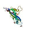 7ad5C  7b76C S: Starting model for refinement C: citing same article ( |
|---|---|
| Similar structure data |


-Links
 PDBj
PDBj- Assembly
| Deposited unit | 
| ||||||||
|---|---|---|---|---|---|---|---|---|---|
| 1 |
| ||||||||
| Unit cell |
|
-Components
| #1: Protein | Mass: 16398.268 Da / Num. of mol.: 1 Source method: isolated from a genetically manipulated source Source: (gene. exp.) Passalora fulva (fungus) / Gene: Ecp11-1 / Production host: Komagataella pastoris (fungus) / References: UniProt: A0A1P8YXI8 | ||||||||
|---|---|---|---|---|---|---|---|---|---|
| #2: Chemical | ChemComp-PEG /   Mass: 106.120 Da / Num. of mol.: 1 / Source method: obtained synthetically / Formula: C4H10O3 Mass: 106.120 Da / Num. of mol.: 1 / Source method: obtained synthetically / Formula: C4H10O3 | ||||||||
| #3: Chemical | ChemComp-GOL /   Mass: 92.094 Da / Num. of mol.: 4 / Source method: obtained synthetically / Formula: C3H8O3 Mass: 92.094 Da / Num. of mol.: 4 / Source method: obtained synthetically / Formula: C3H8O3#4: Chemical | ChemComp-ZN /   Mass: 65.409 Da / Num. of mol.: 4 / Source method: obtained synthetically / Formula: Zn Mass: 65.409 Da / Num. of mol.: 4 / Source method: obtained synthetically / Formula: Zn#5: Water | ChemComp-HOH / |  Mass: 18.015 Da / Num. of mol.: 159 / Source method: isolated from a natural source / Formula: H2O Mass: 18.015 Da / Num. of mol.: 159 / Source method: isolated from a natural source / Formula: H2OHas ligand of interest | N | Has protein modification | Y | |
-Experimental details
-Experiment
| Experiment | Method: X-RAY DIFFRACTION / Number of used crystals: 1 |
|---|
- Sample preparation
| Crystal | Density Matthews: 2.93 Å3/Da / Density % sol: 57.24 % |
|---|---|
| Crystal grow | Temperature: 277 K / Method: vapor diffusion, sitting drop / pH: 6.5 Details: 21% PEG550MME, 10mM Zinc sulfate, 0.1 MES pH6.5, 15% glycerol |
-Data collection
| Diffraction | Mean temperature: 100 K / Serial crystal experiment: N |
|---|---|
| Diffraction source | Source: SYNCHROTRON / Site: SOLEIL / Beamline: PROXIMA 2 / Wavelength: 0.9734 Å |
| Detector | Type: DECTRIS EIGER X 9M / Detector: PIXEL / Date: May 25, 2019 |
| Radiation | Protocol: SINGLE WAVELENGTH / Monochromatic (M) / Laue (L): M / Scattering type: x-ray |
| Radiation wavelength | Wavelength: 0.9734 Å / Relative weight: 1 |
| Reflection | Resolution: 1.62→45.55 Å / Num. obs: 46421 / % possible obs: 99.7 % / Redundancy: 7 % / CC1/2: 0.998 / Rrim(I) all: 0.102 / Net I/σ(I): 10.56 |
| Reflection shell | Resolution: 1.62→1.72 Å / Mean I/σ(I) obs: 0.86 / Num. unique obs: 7420 / CC1/2: 0.604 / Rrim(I) all: 0.142 |
- Processing
| Software |
| ||||||||||||||||||||||||||||||||||||||||||||||||||||||||||||
|---|---|---|---|---|---|---|---|---|---|---|---|---|---|---|---|---|---|---|---|---|---|---|---|---|---|---|---|---|---|---|---|---|---|---|---|---|---|---|---|---|---|---|---|---|---|---|---|---|---|---|---|---|---|---|---|---|---|---|---|---|---|
| Refinement | Method to determine structure: MOLECULAR REPLACEMENT Starting model: 6ZUQ Resolution: 1.62→45.55 Å / Cor.coef. Fo:Fc: 0.969 / Cor.coef. Fo:Fc free: 0.946 / SU B: 2.798 / SU ML: 0.081 / Cross valid method: THROUGHOUT / σ(F): 0 / ESU R: 0.082 / ESU R Free: 0.077 / Stereochemistry target values: MAXIMUM LIKELIHOOD Details: HYDROGENS HAVE BEEN ADDED IN THE RIDING POSITIONS U VALUES : REFINED INDIVIDUALLY
| ||||||||||||||||||||||||||||||||||||||||||||||||||||||||||||
| Solvent computation | Ion probe radii: 0.8 Å / Shrinkage radii: 0.8 Å / VDW probe radii: 1.2 Å / Solvent model: MASK | ||||||||||||||||||||||||||||||||||||||||||||||||||||||||||||
| Displacement parameters | Biso max: 116.68 Å2 / Biso mean: 27.012 Å2 / Biso min: 15.26 Å2
| ||||||||||||||||||||||||||||||||||||||||||||||||||||||||||||
| Refinement step | Cycle: final / Resolution: 1.62→45.55 Å
| ||||||||||||||||||||||||||||||||||||||||||||||||||||||||||||
| Refine LS restraints |
| ||||||||||||||||||||||||||||||||||||||||||||||||||||||||||||
| LS refinement shell | Resolution: 1.62→1.659 Å / Rfactor Rfree error: 0
|
