 Movie
Movie Controller


+ Open data

- Basic information
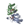
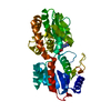
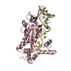
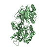
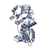
| Entry | Database: PDB / ID: 6zot | ||||||
|---|---|---|---|---|---|---|---|
| Title | Crystal structure of YTHDF3 YTH domain in complex with m6A RNA | ||||||
 Components Components |
| ||||||
 Keywords Keywords | RNA BINDING PROTEIN / YTHDF3 / m6A / complex | ||||||
| Function / homology |  Function and homology information Function and homology informationorganelle assembly / regulation of trophoblast cell migration / N6-methyladenosine-containing RNA reader activity / negative regulation of type I interferon-mediated signaling pathway / mRNA destabilization / positive regulation of translational initiation / regulation of mRNA stability / stress granule assembly / positive regulation of translation / P-body ...organelle assembly / regulation of trophoblast cell migration / N6-methyladenosine-containing RNA reader activity / negative regulation of type I interferon-mediated signaling pathway / mRNA destabilization / positive regulation of translational initiation / regulation of mRNA stability / stress granule assembly / positive regulation of translation / P-body / cytoplasmic stress granule / ribosome binding / mRNA binding / RNA binding / cytoplasm / cytosol Similarity search - Function | ||||||
| Biological species |  Homo sapiens (human) Homo sapiens (human) | ||||||
| Method |  X-RAY DIFFRACTION / SYNCHROTRON / MOLECULAR REPLACEMENT / Resolution: 2.7 Å X-RAY DIFFRACTION / SYNCHROTRON / MOLECULAR REPLACEMENT / Resolution: 2.7 Å | ||||||
 Authors Authors | Bedi, R.K. / Caflisch, A. | ||||||
| Funding support |  Switzerland, 1items Switzerland, 1items
| ||||||
 Citation Citation | Journal: J.Chem.Inf.Model. / Year: 2020 Title: Structural and Dynamic Insights into Redundant Function of YTHDF Proteins. Authors: Li, Y. / Bedi, R.K. / Moroz-Omori, E.V. / Caflisch, A. | ||||||
| History |
|
- Structure visualization
| Structure viewer | Molecule: MolmilJmol/JSmol |
|---|
- Downloads & links
-Download
| PDBx/mmCIF format | 6zot.cif.gz | 104.4 KB | Display | PDBx/mmCIF format |
|---|---|---|---|---|
| PDB format | pdb6zot.ent.gz | 62.9 KB | Display | PDB format |
| PDBx/mmJSON format | 6zot.json.gz | Tree view | PDBx/mmJSON format | |
| Others |  Other downloads Other downloads |
-Validation report
| Arichive directory | https://data.pdbj.org/pub/pdb/validation_reports/zo/6zotftp://data.pdbj.org/pub/pdb/validation_reports/zo/6zot | HTTPS FTP |
|---|
-Related structure data
| Related structure data |  4rciS S: Starting model for refinement |
|---|---|
| Similar structure data |


-Links
 PDBj
PDBj- Assembly
| Deposited unit | 
| ||||||||||||
|---|---|---|---|---|---|---|---|---|---|---|---|---|---|
| 1 |
| ||||||||||||
| Unit cell |
| ||||||||||||
| Components on special symmetry positions |
|
-Components
| #1: Protein | Mass: 23285.250 Da / Num. of mol.: 2 Source method: isolated from a genetically manipulated source Source: (gene. exp.) Homo sapiens (human) / Gene: YTHDF3 / Production host:  #2: RNA chain | | Mass: 1600.035 Da / Num. of mol.: 1 / Source method: obtained synthetically / Source: (synth.) Homo sapiens (human)#3: Water | ChemComp-HOH / |  Mass: 18.015 Da / Num. of mol.: 105 / Source method: isolated from a natural source / Formula: H2O Mass: 18.015 Da / Num. of mol.: 105 / Source method: isolated from a natural source / Formula: H2OHas ligand of interest | Y | |
|---|
-Experimental details
-Experiment
| Experiment | Method: X-RAY DIFFRACTION / Number of used crystals: 1 |
|---|
- Sample preparation
| Crystal | Density Matthews: 1.92 Å3/Da / Density % sol: 35.93 % |
|---|---|
| Crystal grow | Temperature: 295 K / Method: vapor diffusion, hanging drop / Details: 25% PEG 3350, 0.1M Tris pH8, 0.2M NaCl |
-Data collection
| Diffraction | Mean temperature: 100 K / Serial crystal experiment: N |
|---|---|
| Diffraction source | Source: SYNCHROTRON / Site: SLS / Beamline: X06DA / Wavelength: 1 Å |
| Detector | Type: DECTRIS PILATUS 2M-F / Detector: PIXEL / Date: Mar 2, 2019 |
| Radiation | Protocol: SINGLE WAVELENGTH / Monochromatic (M) / Laue (L): M / Scattering type: x-ray |
| Radiation wavelength | Wavelength: 1 Å / Relative weight: 1 |
| Reflection | Resolution: 2.7→45.06 Å / Num. obs: 10844 / % possible obs: 99.8 % / Redundancy: 14.95 % / Biso Wilson estimate: 36.27 Å2 / CC1/2: 0.997 / Net I/σ(I): 7.25 |
| Reflection shell | Resolution: 2.7→2.86 Å / Num. unique obs: 1696 / CC1/2: 0.617 |
- Processing
| Software |
| |||||||||||||||||||||||||||||||||||
|---|---|---|---|---|---|---|---|---|---|---|---|---|---|---|---|---|---|---|---|---|---|---|---|---|---|---|---|---|---|---|---|---|---|---|---|---|
| Refinement | Method to determine structure: MOLECULAR REPLACEMENT Starting model: 4RCI Resolution: 2.7→45.06 Å / SU ML: 0.4001 / Cross valid method: FREE R-VALUE / σ(F): 1.36 / Phase error: 29.9496 Stereochemistry target values: GeoStd + Monomer Library + CDL v1.2
| |||||||||||||||||||||||||||||||||||
| Solvent computation | Shrinkage radii: 0.9 Å / VDW probe radii: 1.11 Å / Solvent model: FLAT BULK SOLVENT MODEL | |||||||||||||||||||||||||||||||||||
| Displacement parameters | Biso mean: 29.55 Å2 | |||||||||||||||||||||||||||||||||||
| Refinement step | Cycle: LAST / Resolution: 2.7→45.06 Å
| |||||||||||||||||||||||||||||||||||
| Refine LS restraints |
| |||||||||||||||||||||||||||||||||||
| LS refinement shell |
|
