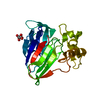
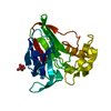
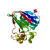
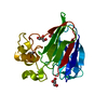
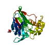
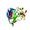
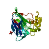
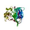
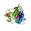
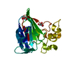
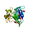
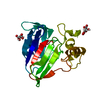
- PDB-6zhn: 3D electron diffraction structure of thaumatin from Thaumatococcu... -
+
データを開く
IDまたはキーワード:
読み込み中...
-
基本情報
登録情報
データベース: PDB / ID: 6zhn
タイトル
3D electron diffraction structure of thaumatin from Thaumatococcus daniellii
要素
Thaumatin-1
キーワード
PLANT PROTEIN / nanocrystals
機能・相同性
Thaumatin, conserved site / Thaumatin family signature. / Thaumatin family / Thaumatin family / Thaumatin family profile. / Thaumatin family / Osmotin/thaumatin-like superfamily / cytoplasmic vesicle / Thaumatin I
ジャーナル: Acta Crystallogr D Struct Biol / 年: 2021 タイトル: Statistically correcting dynamical electron scattering improves the refinement of protein nanocrystals, including charge refinement of coordinated metals. 著者: Thorsten B Blum / Dominique Housset / Max T B Clabbers / Eric van Genderen / Maria Bacia-Verloop / Ulrich Zander / Andrew A McCarthy / Guy Schoehn / Wai Li Ling / Jan Pieter Abrahams / 要旨: Electron diffraction allows protein structure determination when only nanosized crystals are available. Nevertheless, multiple elastic (or dynamical) scattering, which is prominent in electron ...Electron diffraction allows protein structure determination when only nanosized crystals are available. Nevertheless, multiple elastic (or dynamical) scattering, which is prominent in electron diffraction, is a concern. Current methods for modeling dynamical scattering by multi-slice or Bloch wave approaches are not suitable for protein crystals because they are not designed to cope with large molecules. Here, dynamical scattering of nanocrystals of insulin, thermolysin and thaumatin was limited by collecting data from thin crystals. To accurately measure the weak diffraction signal from the few unit cells in the thin crystals, a low-noise hybrid pixel Timepix electron-counting detector was used. The remaining dynamical component was further reduced in refinement using a likelihood-based correction, which was introduced previously for analyzing electron diffraction data of small-molecule nanocrystals and was adapted here for protein crystals. The procedure is shown to notably improve the structural refinement, in one case allowing the location of solvent molecules. It also allowed refinement of the charge states of bound metal atoms, an important element in protein function, through B-factor analysis of the metal atoms and their ligands. These results clearly increase the value of macromolecular electron crystallography as a complementary structural biology technique.
解像度: 2.76→45.69 Å / Cor.coef. Fo:Fc: 0.836 / Cor.coef. Fo:Fc free: 0.787 / SU B: 16.607 / SU ML: 0.37 / 交差検証法: THROUGHOUT / σ(F): 0 / ESU R Free: 0.619 / 立体化学のターゲット値: MAXIMUM LIKELIHOOD 詳細: HYDROGENS HAVE BEEN ADDED IN THE RIDING POSITIONS U VALUES : REFINED INDIVIDUALLY
Rfactor
反射数
%反射
Selection details
Rfree
0.321
231
5 %
RANDOM
Rwork
0.2801
-
-
-
obs
0.2822
4364
65.61 %
-
溶媒の処理
イオンプローブ半径: 0.8 Å / 減衰半径: 0.8 Å / VDWプローブ半径: 1.2 Å / 溶媒モデル: MASK
 ムービー
ムービー コントローラー


 データを開く
 基本情報
基本情報 要素
要素 キーワード
キーワード 機能・相同性情報
 Thaumatococcus daniellii (植物)
 分子置換 / クライオ電子顕微鏡法 / 解像度: 2.76 Å
  データ登録者
 フランス,
フランス,  スイス, 4件
  引用
引用 構造の表示
構造の表示 ダウンロードとリンク
 その他のダウンロード
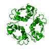


 PDBj
PDBj
 集合体
集合体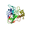

 分子量: 35.453 Da / 分子数: 1 / 由来タイプ: 合成 / 式: Cl
 試料調製
試料調製
 解析
解析