- PDB-6zhb: 3D electron diffraction structure of bovine insulin -
+
Open data
ID or keywords:
Loading...
-
Basic information
Entry
Database: PDB / ID: 6zhb
Title
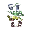
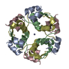
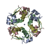
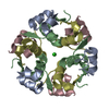
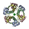
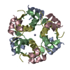
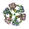
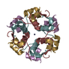
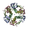
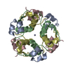
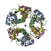
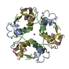
3D electron diffraction structure of bovine insulin
Components
(Insulin) x 2
Keywords
HORMONE / INSULIN FAMILY / CARBOHYDRATE METABOLISM / HORMONE-GROWTH
Function / homology
Function and homology information
estradiol secretion / positive regulation of blood circulation / negative regulation of lactation / glucose import in response to insulin stimulus / positive regulation of cell maturation / response to L-arginine / positive regulation of mammary gland epithelial cell proliferation / positive regulation of lactation / negative regulation of appetite / response to butyrate ...estradiol secretion / positive regulation of blood circulation / negative regulation of lactation / glucose import in response to insulin stimulus / positive regulation of cell maturation / response to L-arginine / positive regulation of mammary gland epithelial cell proliferation / positive regulation of lactation / negative regulation of appetite / response to butyrate / feeding behavior / response to growth hormone / positive regulation of peptide hormone secretion / response to food / positive regulation of Rho protein signal transduction / protein secretion / negative regulation of lipid catabolic process / response to glucose / insulin receptor binding / positive regulation of protein secretion / hormone activity / response to nutrient levels / glucose metabolic process / positive regulation of insulin secretion / glucose homeostasis / response to heat / positive regulation of phosphatidylinositol 3-kinase/protein kinase B signal transduction / positive regulation of gene expression / negative regulation of apoptotic process / : / identical protein binding Similarity search - Function
Insulin / Insulin family / Insulin-like / Insulin/IGF/Relaxin family / Insulin / insulin-like growth factor / relaxin family. / Insulin, conserved site / Insulin family signature. / Insulin-like superfamily Similarity search - Domain/homology
Journal: Acta Crystallogr D Struct Biol / Year: 2021 Title: Statistically correcting dynamical electron scattering improves the refinement of protein nanocrystals, including charge refinement of coordinated metals. Authors: Thorsten B Blum / Dominique Housset / Max T B Clabbers / Eric van Genderen / Maria Bacia-Verloop / Ulrich Zander / Andrew A McCarthy / Guy Schoehn / Wai Li Ling / Jan Pieter Abrahams / Abstract: Electron diffraction allows protein structure determination when only nanosized crystals are available. Nevertheless, multiple elastic (or dynamical) scattering, which is prominent in electron ...Electron diffraction allows protein structure determination when only nanosized crystals are available. Nevertheless, multiple elastic (or dynamical) scattering, which is prominent in electron diffraction, is a concern. Current methods for modeling dynamical scattering by multi-slice or Bloch wave approaches are not suitable for protein crystals because they are not designed to cope with large molecules. Here, dynamical scattering of nanocrystals of insulin, thermolysin and thaumatin was limited by collecting data from thin crystals. To accurately measure the weak diffraction signal from the few unit cells in the thin crystals, a low-noise hybrid pixel Timepix electron-counting detector was used. The remaining dynamical component was further reduced in refinement using a likelihood-based correction, which was introduced previously for analyzing electron diffraction data of small-molecule nanocrystals and was adapted here for protein crystals. The procedure is shown to notably improve the structural refinement, in one case allowing the location of solvent molecules. It also allowed refinement of the charge states of bound metal atoms, an important element in protein function, through B-factor analysis of the metal atoms and their ligands. These results clearly increase the value of macromolecular electron crystallography as a complementary structural biology technique.
Resolution: 3.25→30.3 Å / Cor.coef. Fo:Fc: 0.934 / Cor.coef. Fo:Fc free: 0.754 / SU B: 58.278 / SU ML: 0.899 / Cross valid method: THROUGHOUT / σ(F): 0 / ESU R Free: 0.989 / Stereochemistry target values: MAXIMUM LIKELIHOOD Details: HYDROGENS HAVE BEEN ADDED IN THE RIDING POSITIONS U VALUES : REFINED INDIVIDUALLY
Rfactor
Num. reflection
% reflection
Selection details
Rfree
0.3189
108
9.6 %
RANDOM
Rwork
0.1809
-
-
-
obs
0.1942
1019
84.36 %
-
Solvent computation
Ion probe radii: 0.8 Å / Shrinkage radii: 0.8 Å / VDW probe radii: 1.2 Å / Solvent model: MASK
In the structure databanks used in Yorodumi, some data are registered as the other names, "COVID-19 virus" and "2019-nCoV". Here are the details of the virus and the list of structure data.
Jan 31, 2019. EMDB accession codes are about to change! (news from PDBe EMDB page)
EMDB accession codes are about to change! (news from PDBe EMDB page)
The allocation of 4 digits for EMDB accession codes will soon come to an end. Whilst these codes will remain in use, new EMDB accession codes will include an additional digit and will expand incrementally as the available range of codes is exhausted. The current 4-digit format prefixed with “EMD-” (i.e. EMD-XXXX) will advance to a 5-digit format (i.e. EMD-XXXXX), and so on. It is currently estimated that the 4-digit codes will be depleted around Spring 2019, at which point the 5-digit format will come into force.
The EM Navigator/Yorodumi systems omit the EMD- prefix.
Related info.:Q: What is EMD? / ID/Accession-code notation in Yorodumi/EM Navigator
Yorodumi is a browser for structure data from EMDB, PDB, SASBDB, etc.
This page is also the successor to EM Navigator detail page, and also detail information page/front-end page for Omokage search.
The word "yorodu" (or yorozu) is an old Japanese word meaning "ten thousand". "mi" (miru) is to see.
Related info.:EMDB / PDB / SASBDB / Comparison of 3 databanks / Yorodumi Search / Aug 31, 2016. New EM Navigator & Yorodumi / Yorodumi Papers / Jmol/JSmol / Function and homology information / Changes in new EM Navigator and Yorodumi
 Movie
Movie Controller


 Open data
 Basic information
 Components
 Keywords
 Function and homology information
 MOLECULAR REPLACEMENT / cryo EM / Resolution: 3.25 Å
  Authors
 France,
  Switzerland, 4items
  Citation
 Structure visualization
 Downloads & links
 Other downloads


 PDBj
PDBj


 Assembly

 Mass: 65.409 Da / Num. of mol.: 1 / Source method: obtained synthetically / Formula: Zn
 Sample preparation
 Processing