 Movie
Movie Controller


+ Open data

- Basic information
| Entry | Database: PDB / ID: 2a3g | ||||||
|---|---|---|---|---|---|---|---|
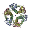
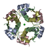
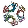
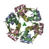
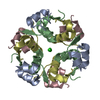
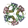
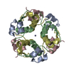
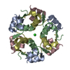
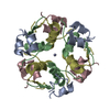
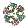
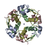
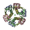
| Title | The structure of T6 bovine insulin | ||||||
 Components Components | (Insulin) x 2 | ||||||
 Keywords Keywords | HORMONE/GROWTH FACTOR / hormone / insulin family / carbohydrate metabolism / HORMONE-GROWTH FACTOR COMPLEX | ||||||
| Function / homology |  Function and homology information Function and homology informationestradiol secretion / positive regulation of blood circulation / negative regulation of lactation / glucose import in response to insulin stimulus / positive regulation of cell maturation / response to L-arginine / positive regulation of mammary gland epithelial cell proliferation / positive regulation of lactation / response to butyrate / negative regulation of appetite ...estradiol secretion / positive regulation of blood circulation / negative regulation of lactation / glucose import in response to insulin stimulus / positive regulation of cell maturation / response to L-arginine / positive regulation of mammary gland epithelial cell proliferation / positive regulation of lactation / response to butyrate / negative regulation of appetite / feeding behavior / response to growth hormone / positive regulation of peptide hormone secretion / response to food / positive regulation of Rho protein signal transduction / protein secretion / negative regulation of lipid catabolic process / response to glucose / insulin receptor binding / positive regulation of protein secretion / hormone activity / response to nutrient levels / glucose metabolic process / positive regulation of insulin secretion / glucose homeostasis / response to heat / positive regulation of phosphatidylinositol 3-kinase/protein kinase B signal transduction / positive regulation of gene expression / negative regulation of apoptotic process / : / identical protein binding Similarity search - Function | ||||||
| Biological species |  | ||||||
| Method |  X-RAY DIFFRACTION / MOLECULAR REPLACEMENT / Resolution: 2.25 Å X-RAY DIFFRACTION / MOLECULAR REPLACEMENT / Resolution: 2.25 Å | ||||||
 Authors Authors | Smith, G.D. / Pangborn, W.A. / Blessing, R.H. | ||||||
 Citation Citation | Journal: Acta Crystallogr.,Sect.D / Year: 2005 Title: The structure of T6 bovine insulin. Authors: Smith, G.D. / Pangborn, W.A. / Blessing, R.H. #1: Journal: Philos.Trans.R.Soc.London,Ser.B / Year: 1988Title: The structure of 2Zn pig insulin crystals at 1.5 A resolution Authors: Baker, E.N. / Blundell, T.L. / Cutfield, J.F. / Cutfield, S.M. / Dodson, E.J. / Dodson, G.G. / Hodgkin, D.M. / Hubbard, R.E. / Issacs, N.W. / Reynolds, C.D. / Sakabe, K. / Sakabe, N. / Vijayan, N.M. #2: Journal: Acta Crystallogr.,Sect.D / Year: 2003Title: The structure of T6 human insulin at 1.0 A resolution Authors: Smith, G.D. / Pangborn, W.A. / Blessing, R.H. #3: Journal: Acta Crystallogr.,Sect.D / Year: 2003Title: Lessons from an aged, dried crystal of T(6) human insulin Authors: Smith, G.D. / Blessing, R.H. | ||||||
| History |
|
- Structure visualization
| Structure viewer | Molecule: MolmilJmol/JSmol |
|---|
- Downloads & links
-Download
| PDBx/mmCIF format | 2a3g.cif.gz | 34.3 KB | Display | PDBx/mmCIF format |
|---|---|---|---|---|
| PDB format | pdb2a3g.ent.gz | 23.2 KB | Display | PDB format |
| PDBx/mmJSON format | 2a3g.json.gz | Tree view | PDBx/mmJSON format | |
| Others |  Other downloads Other downloads |
-Validation report
| Arichive directory | https://data.pdbj.org/pub/pdb/validation_reports/a3/2a3gftp://data.pdbj.org/pub/pdb/validation_reports/a3/2a3g | HTTPS FTP |
|---|
-Related structure data
| Related structure data | 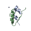 4insS S: Starting model for refinement |
|---|---|
| Similar structure data |

-Links
 PDBj
PDBj


- Assembly
| Deposited unit | 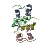
| ||||||||||||||||||||||||
|---|---|---|---|---|---|---|---|---|---|---|---|---|---|---|---|---|---|---|---|---|---|---|---|---|---|
| 1 | 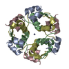
| ||||||||||||||||||||||||
| Unit cell |
| ||||||||||||||||||||||||
| Components on special symmetry positions |
| ||||||||||||||||||||||||
| Details | THE SECOND PART OF THE BIOLOGICAL ASSEMBLY IS GENERATED BY THE THREE FOLD AXIS: -Y, X-Y, Z / THE THIRD PART OF THE BIOLOGICAL ASSEMBLY IS GENERATED BY THE THREE FOLD AXIS: Y-X, -X, Z |
-Components
| #1: Protein/peptide | Mass: 2339.645 Da / Num. of mol.: 2 / Fragment: insulin A chain, residues 85-105 / Source method: isolated from a natural source / Source: (natural) #2: Protein/peptide | Mass: 3403.927 Da / Num. of mol.: 2 / Fragment: insulin B chain, residues 25-54 / Source method: isolated from a natural source / Source: (natural) #3: Chemical |   Mass: 65.409 Da / Num. of mol.: 2 / Source method: obtained synthetically / Formula: Zn Mass: 65.409 Da / Num. of mol.: 2 / Source method: obtained synthetically / Formula: Zn#4: Water | ChemComp-HOH / |  Mass: 18.015 Da / Num. of mol.: 56 / Source method: isolated from a natural source / Formula: H2O Mass: 18.015 Da / Num. of mol.: 56 / Source method: isolated from a natural source / Formula: H2OHas protein modification | Y | |
|---|
-Experimental details
-Experiment
| Experiment | Method: X-RAY DIFFRACTION / Number of used crystals: 1 |
|---|
- Sample preparation
| Crystal | Density Matthews: 1.9 Å3/Da / Density % sol: 35.2 % |
|---|---|
| Crystal grow | Temperature: 298 K / pH: 6.2 Details: HCl, ZINC ACETATE, SODIUM CITRATE, ACETONE, pH 6.2, SLOW COOLING, temperature 298 K, pH 6.20 |
-Data collection
| Diffraction | Mean temperature: 298 K |
|---|---|
| Diffraction source | Source: ROTATING ANODE / Type: RIGAKU RU200 / Wavelength: 1.54178 |
| Detector | Type: RIGAKU RAXIS IIC / Detector: IMAGE PLATE / Date: Aug 11, 1997 / Details: GRAPHITE MONOCHROMETER |
| Radiation | Monochromator: GRAPHITE / Protocol: SINGLE WAVELENGTH / Monochromatic (M) / Laue (L): M / Scattering type: x-ray |
| Radiation wavelength | Wavelength: 1.54178 Å / Relative weight: 1 |
| Reflection | Resolution: 2.2→30.5 Å / Num. obs: 4345 / % possible obs: 99.5 % / Observed criterion σ(I): 0 / Redundancy: 4.04 % / Biso Wilson estimate: 25.9 Å2 / Rmerge(I) obs: 0.051 / Net I/σ(I): 18.1 |
| Reflection shell | Resolution: 2.2→2.25 Å / Rmerge(I) obs: 0.207 / Mean I/σ(I) obs: 2.81 / % possible all: 94 |
- Processing
| Software |
| ||||||||||||||||||||||||||||||||||||||||||||||||||||||||||||||||||||||||||||||||
|---|---|---|---|---|---|---|---|---|---|---|---|---|---|---|---|---|---|---|---|---|---|---|---|---|---|---|---|---|---|---|---|---|---|---|---|---|---|---|---|---|---|---|---|---|---|---|---|---|---|---|---|---|---|---|---|---|---|---|---|---|---|---|---|---|---|---|---|---|---|---|---|---|---|---|---|---|---|---|---|---|---|
| Refinement | Method to determine structure: MOLECULAR REPLACEMENT Starting model: PDB ENTRY 4INS Resolution: 2.25→30.5 Å / Isotropic thermal model: ISOTROPIC / Cross valid method: THROUGHOUT / σ(F): 0 / Stereochemistry target values: ENGH & HUBER
| ||||||||||||||||||||||||||||||||||||||||||||||||||||||||||||||||||||||||||||||||
| Solvent computation | Solvent model: FLAT MODEL / Bsol: 38.3 Å2 / ksol: 0.29 e/Å3 | ||||||||||||||||||||||||||||||||||||||||||||||||||||||||||||||||||||||||||||||||
| Displacement parameters | Biso mean: 29.4 Å2 | ||||||||||||||||||||||||||||||||||||||||||||||||||||||||||||||||||||||||||||||||
| Refine analyze |
| ||||||||||||||||||||||||||||||||||||||||||||||||||||||||||||||||||||||||||||||||
| Refinement step | Cycle: LAST / Resolution: 2.25→30.5 Å
| ||||||||||||||||||||||||||||||||||||||||||||||||||||||||||||||||||||||||||||||||
| Refine LS restraints |
| ||||||||||||||||||||||||||||||||||||||||||||||||||||||||||||||||||||||||||||||||
| LS refinement shell | Resolution: 2.25→2.39 Å / Rfactor Rfree error: 0.032
|
