- PDB-6zhb: 3D electron diffraction structure of bovine insulin -
+
データを開く
IDまたはキーワード:
読み込み中...
-
基本情報
登録情報
データベース: PDB / ID: 6zhb
タイトル
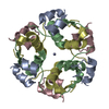
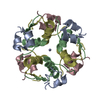
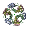
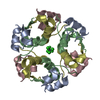
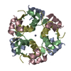
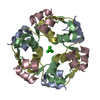
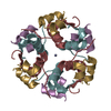
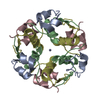
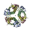
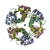
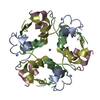
3D electron diffraction structure of bovine insulin
要素
(Insulin) x 2
キーワード
HORMONE / INSULIN FAMILY / CARBOHYDRATE METABOLISM / HORMONE-GROWTH
機能・相同性
機能・相同性情報
estradiol secretion / positive regulation of blood circulation / negative regulation of lactation / glucose import in response to insulin stimulus / positive regulation of cell maturation / positive regulation of lactation / response to L-arginine / positive regulation of mammary gland epithelial cell proliferation / response to butyrate / negative regulation of appetite ...estradiol secretion / positive regulation of blood circulation / negative regulation of lactation / glucose import in response to insulin stimulus / positive regulation of cell maturation / positive regulation of lactation / response to L-arginine / positive regulation of mammary gland epithelial cell proliferation / response to butyrate / negative regulation of appetite / feeding behavior / response to growth hormone / response to food / positive regulation of peptide hormone secretion / positive regulation of Rho protein signal transduction / protein secretion / response to glucose / negative regulation of lipid catabolic process / positive regulation of protein secretion / insulin receptor binding / response to nutrient levels / hormone activity / positive regulation of insulin secretion / glucose metabolic process / glucose homeostasis / response to heat / positive regulation of phosphatidylinositol 3-kinase/protein kinase B signal transduction / positive regulation of gene expression / negative regulation of apoptotic process / extracellular space / identical protein binding 類似検索 - 分子機能
Insulin / Insulin family / Insulin-like / Insulin/IGF/Relaxin family / Insulin / insulin-like growth factor / relaxin family. / Insulin, conserved site / Insulin family signature. / Insulin-like superfamily 類似検索 - ドメイン・相同性
ジャーナル: Acta Crystallogr D Struct Biol / 年: 2021 タイトル: Statistically correcting dynamical electron scattering improves the refinement of protein nanocrystals, including charge refinement of coordinated metals. 著者: Thorsten B Blum / Dominique Housset / Max T B Clabbers / Eric van Genderen / Maria Bacia-Verloop / Ulrich Zander / Andrew A McCarthy / Guy Schoehn / Wai Li Ling / Jan Pieter Abrahams / 要旨: Electron diffraction allows protein structure determination when only nanosized crystals are available. Nevertheless, multiple elastic (or dynamical) scattering, which is prominent in electron ...Electron diffraction allows protein structure determination when only nanosized crystals are available. Nevertheless, multiple elastic (or dynamical) scattering, which is prominent in electron diffraction, is a concern. Current methods for modeling dynamical scattering by multi-slice or Bloch wave approaches are not suitable for protein crystals because they are not designed to cope with large molecules. Here, dynamical scattering of nanocrystals of insulin, thermolysin and thaumatin was limited by collecting data from thin crystals. To accurately measure the weak diffraction signal from the few unit cells in the thin crystals, a low-noise hybrid pixel Timepix electron-counting detector was used. The remaining dynamical component was further reduced in refinement using a likelihood-based correction, which was introduced previously for analyzing electron diffraction data of small-molecule nanocrystals and was adapted here for protein crystals. The procedure is shown to notably improve the structural refinement, in one case allowing the location of solvent molecules. It also allowed refinement of the charge states of bound metal atoms, an important element in protein function, through B-factor analysis of the metal atoms and their ligands. These results clearly increase the value of macromolecular electron crystallography as a complementary structural biology technique.
解像度: 3.25→30.3 Å / Cor.coef. Fo:Fc: 0.934 / Cor.coef. Fo:Fc free: 0.754 / SU B: 58.278 / SU ML: 0.899 / 交差検証法: THROUGHOUT / σ(F): 0 / ESU R Free: 0.989 / 立体化学のターゲット値: MAXIMUM LIKELIHOOD 詳細: HYDROGENS HAVE BEEN ADDED IN THE RIDING POSITIONS U VALUES : REFINED INDIVIDUALLY
Rfactor
反射数
%反射
Selection details
Rfree
0.3189
108
9.6 %
RANDOM
Rwork
0.1809
-
-
-
obs
0.1942
1019
84.36 %
-
溶媒の処理
イオンプローブ半径: 0.8 Å / 減衰半径: 0.8 Å / VDWプローブ半径: 1.2 Å / 溶媒モデル: MASK
 ムービー
ムービー コントローラー


 データを開く
 基本情報
基本情報 要素
要素 キーワード
キーワード 機能・相同性情報
 分子置換 / クライオ電子顕微鏡法 / 解像度: 3.25 Å
  データ登録者
 フランス,
フランス,  スイス, 4件
  引用
引用 構造の表示
構造の表示 ダウンロードとリンク
 その他のダウンロード


 PDBj
PDBj


 集合体
集合体

 分子量: 65.409 Da / 分子数: 1 / 由来タイプ: 合成 / 式: Zn
 試料調製
試料調製
 解析
解析