HLA-A specific inhibitory MHC class I receptor activity / : / negative regulation of serotonin secretion / MHC class Ib protein complex binding / HLA-B specific inhibitory MHC class I receptor activity / immune response-inhibiting cell surface receptor signaling pathway / inhibitory MHC class I receptor activity / antigenic variation / negative regulation of dendritic cell differentiation / Fc receptor mediated inhibitory signaling pathway ...HLA-A specific inhibitory MHC class I receptor activity / : / negative regulation of serotonin secretion / MHC class Ib protein complex binding / HLA-B specific inhibitory MHC class I receptor activity / immune response-inhibiting cell surface receptor signaling pathway / inhibitory MHC class I receptor activity / antigenic variation / negative regulation of dendritic cell differentiation / Fc receptor mediated inhibitory signaling pathway / MHC class Ib receptor activity / MHC class Ib protein binding / negative regulation of T cell mediated cytotoxicity / negative regulation of CD8-positive, alpha-beta T cell activation / immune response-regulating signaling pathway / negative regulation of transforming growth factor beta production / MHC class I receptor activity / negative regulation of cytokine production involved in immune response / dendritic cell differentiation / interleukin-10-mediated signaling pathway / negative regulation of osteoclast development / negative regulation of T cell activation via T cell receptor contact with antigen bound to MHC molecule on antigen presenting cell / protein phosphatase 1 binding / negative regulation of interleukin-12 production / negative regulation of alpha-beta T cell activation / negative regulation of dendritic cell apoptotic process / negative regulation of endocytosis / negative regulation of interferon-beta production / negative regulation of mononuclear cell proliferation / negative regulation of natural killer cell mediated cytotoxicity / T cell proliferation involved in immune response / positive regulation of macrophage cytokine production / negative regulation of interleukin-10 production / negative regulation of type II interferon production / negative regulation of calcium ion transport / negative regulation of cell cycle / negative regulation of tumor necrosis factor production / MHC class I protein binding / negative regulation of T cell proliferation / positive regulation of defense response to virus by host / SH2 domain binding / receptor internalization / positive regulation of type II interferon production / response to virus / Immunoregulatory interactions between a Lymphoid and a non-Lymphoid cell / cellular response to lipopolysaccharide / defense response to virus / adaptive immune response / cell surface receptor signaling pathway / positive regulation of apoptotic process / external side of plasma membrane / positive regulation of gene expression / host cell plasma membrane / signal transduction / protein homodimerization activity / positive regulation of transcription by RNA polymerase II / extracellular region / membrane / plasma membrane / cytoplasm Similarity search - Function
In the structure databanks used in Yorodumi, some data are registered as the other names, "COVID-19 virus" and "2019-nCoV". Here are the details of the virus and the list of structure data.
Jan 31, 2019. EMDB accession codes are about to change! (news from PDBe EMDB page)
EMDB accession codes are about to change! (news from PDBe EMDB page)
The allocation of 4 digits for EMDB accession codes will soon come to an end. Whilst these codes will remain in use, new EMDB accession codes will include an additional digit and will expand incrementally as the available range of codes is exhausted. The current 4-digit format prefixed with “EMD-” (i.e. EMD-XXXX) will advance to a 5-digit format (i.e. EMD-XXXXX), and so on. It is currently estimated that the 4-digit codes will be depleted around Spring 2019, at which point the 5-digit format will come into force.
The EM Navigator/Yorodumi systems omit the EMD- prefix.
Related info.:Q: What is EMD? / ID/Accession-code notation in Yorodumi/EM Navigator
Yorodumi is a browser for structure data from EMDB, PDB, SASBDB, etc.
This page is also the successor to EM Navigator detail page, and also detail information page/front-end page for Omokage search.
The word "yorodu" (or yorozu) is an old Japanese word meaning "ten thousand". "mi" (miru) is to see.
Related info.:EMDB / PDB / SASBDB / Comparison of 3 databanks / Yorodumi Search / Aug 31, 2016. New EM Navigator & Yorodumi / Yorodumi Papers / Jmol/JSmol / Function and homology information / Changes in new EM Navigator and Yorodumi
 Movie
Movie Controller


 Open data
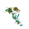
 Basic information
 Components
 Keywords
 Function and homology information
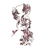
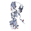
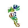
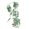
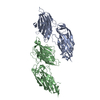
 Homo sapiens (human)
 X-RAY DIFFRACTION /
  Authors
 Citation
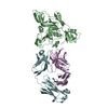
 Structure visualization
 Downloads & links
 Other downloads
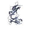


 PDBj
PDBj


 Assembly
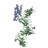


 Type: D-saccharide, beta linking / Mass: 221.208 Da / Num. of mol.: 2
 Sample preparation
 / Beamline: PROXIMA 1 / Wavelength: 0.97857 Å
 Processing