[English] 日本語
Yorodumi
- PDB-6z6e: Crystal structure of the HK97 bacteriophage small terminase -

+
Open data


ID or keywords:

Loading...

-
Basic information

Entry
Database: PDB / ID: 6z6e
TitleCrystal structure of the HK97 bacteriophage small terminase
ComponentsTerminase small subunit
KeywordsVIRAL PROTEIN / genome packaging / bacteriophage / DNA binding
Function / homologyIODIDE ION / Terminase small subunit
Function and homology information
Biological speciesEnterobacteria phage HK97 (virus)
MethodX-RAY DIFFRACTION / SYNCHROTRON / SAD / Resolution: 1.4 Å
AuthorsFung, H.K.H. / Chechik, M. / Baumann, C.G. / Antson, A.A.
Funding support United Kingdom, 2items
OrganizationGrant numberCountry
Wellcome Trust098230 United Kingdom
Wellcome Trust095024MA United Kingdom
Citation
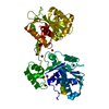
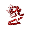
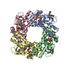
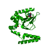
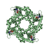
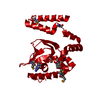
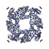
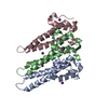
Journal: Nucleic Acids Res / Year: 2022
Title: Structural basis of DNA packaging by a ring-type ATPase from an archetypal viral system.
Authors: Herman K H Fung / Shelley Grimes / Alexis Huet / Robert L Duda / Maria Chechik / Joseph Gault / Carol V Robinson / Roger W Hendrix / Paul J Jardine / James F Conway / Christoph G Baumann / Alfred A Antson /
Abstract: Many essential cellular processes rely on substrate rotation or translocation by a multi-subunit, ring-type NTPase. A large number of double-stranded DNA viruses, including tailed bacteriophages and ...Many essential cellular processes rely on substrate rotation or translocation by a multi-subunit, ring-type NTPase. A large number of double-stranded DNA viruses, including tailed bacteriophages and herpes viruses, use a homomeric ring ATPase to processively translocate viral genomic DNA into procapsids during assembly. Our current understanding of viral DNA packaging comes from three archetypal bacteriophage systems: cos, pac and phi29. Detailed mechanistic understanding exists for pac and phi29, but not for cos. Here, we reconstituted in vitro a cos packaging system based on bacteriophage HK97 and provided a detailed biochemical and structural description. We used a photobleaching-based, single-molecule assay to determine the stoichiometry of the DNA-translocating ATPase large terminase. Crystal structures of the large terminase and DNA-recruiting small terminase, a first for a biochemically defined cos system, reveal mechanistic similarities between cos and pac systems. At the same time, mutational and biochemical analyses indicate a new regulatory mechanism for ATPase multimerization and coordination in the HK97 system. This work therefore establishes a framework for studying the evolutionary relationships between ATP-dependent DNA translocation machineries in double-stranded DNA viruses.
History
DepositionMay 28, 2020Deposition site: PDBE / Processing site: PDBE
Revision 1.0Jun 9, 2021Provider: repository / Type: Initial release
Revision 1.1Dec 21, 2022Group: Database references / Category: citation / citation_author / database_2
Item: _citation.country / _citation.journal_abbrev ..._citation.country / _citation.journal_abbrev / _citation.journal_id_ASTM / _citation.journal_id_CSD / _citation.journal_id_ISSN / _citation.journal_volume / _citation.page_first / _citation.page_last / _citation.pdbx_database_id_DOI / _citation.pdbx_database_id_PubMed / _citation.title / _citation.year / _citation_author.identifier_ORCID / _database_2.pdbx_DOI / _database_2.pdbx_database_accession
Revision 1.2Jun 19, 2024Group: Data collection / Refinement description
Category: chem_comp_atom / chem_comp_bond / struct_ncs_dom_lim
Item: _struct_ncs_dom_lim.beg_auth_comp_id / _struct_ncs_dom_lim.beg_label_asym_id ..._struct_ncs_dom_lim.beg_auth_comp_id / _struct_ncs_dom_lim.beg_label_asym_id / _struct_ncs_dom_lim.beg_label_comp_id / _struct_ncs_dom_lim.beg_label_seq_id / _struct_ncs_dom_lim.end_auth_comp_id / _struct_ncs_dom_lim.end_label_asym_id / _struct_ncs_dom_lim.end_label_comp_id / _struct_ncs_dom_lim.end_label_seq_id

-
Structure visualization

Structure viewerMolecule:
MolmilJmol/JSmol

Downloads & links

-
Assembly

Deposited unit
A: Terminase small subunit
B: Terminase small subunit
C: Terminase small subunit
hetero molecules


Theoretical massNumber of molelcules
Total (without water)56,24112
Polymers55,0993
Non-polymers1,1429
Water6,089338
1
A: Terminase small subunit
B: Terminase small subunit
C: Terminase small subunit
hetero molecules

A: Terminase small subunit
B: Terminase small subunit
C: Terminase small subunit
hetero molecules

A: Terminase small subunit
B: Terminase small subunit
C: Terminase small subunit
hetero molecules


Theoretical massNumber of molelcules
Total (without water)168,72436
Polymers165,2989
Non-polymers3,42627
Water1629
TypeNameSymmetry operationNumber
identity operation1_555x,y,z1
crystal symmetry operation2_555-y,x-y,z1
crystal symmetry operation3_555-x+y,-x,z1
Buried area27010 Å2
ΔGint-44 kcal/mol
Surface area37340 Å2
MethodPISA
Unit cell
Length a, b, c (Å)123.281, 123.281, 78.991
Angle α, β, γ (deg.)90.000, 90.000, 120.000
Int Tables number146
Space group name H-MH3
Noncrystallographic symmetry (NCS)NCS domain:
IDEns-IDDetails
11A
21B
12A
22C
13B
23C

NCS domain segments:

Component-ID: _ / End auth comp-ID: THR / End label comp-ID: THR / Refine code: _

Dom-IDEns-IDBeg auth comp-IDBeg label comp-IDAuth asym-IDLabel asym-IDAuth seq-IDLabel seq-ID
11VALVALAA23 - 12322 - 122
21VALVALBB23 - 12322 - 122
12ASPASPAA24 - 12323 - 122
22ASPASPCC24 - 12323 - 122
13ASPASPBB24 - 12323 - 122
23ASPASPCC24 - 12323 - 122

NCS ensembles :
ID
1
2
3

-
Components

#1: Protein Terminase small subunit


Mass: 18366.453 Da / Num. of mol.: 3
Source method: isolated from a genetically manipulated source
Source: (gene. exp.) Enterobacteria phage HK97 (virus) / Production host: Escherichia coli (E. coli) / References: UniProt: Q9MBW4
#2: Chemical
ChemComp-IOD / IODIDE ION


Mass: 126.904 Da / Num. of mol.: 9 / Source method: obtained synthetically / Formula: I
#3: Water ChemComp-HOH / water


Mass: 18.015 Da / Num. of mol.: 338 / Source method: isolated from a natural source / Formula: H2O
Has ligand of interestN

-
Experimental details

-
Experiment

ExperimentMethod: X-RAY DIFFRACTION / Number of used crystals: 1

-
Sample preparation

CrystalDensity Matthews: 2.1 Å3/Da / Density % sol: 41.33 %
Crystal growTemperature: 293 K / Method: vapor diffusion, hanging drop / Details: 9% (w/v) PEG 3350, 0.1 M succinic acid, pH 7.0

-
Data collection

DiffractionMean temperature: 100 K / Serial crystal experiment: N
Diffraction sourceSource: SYNCHROTRON / Site: Diamond / Beamline: I02 / Wavelength: 0.9763 Å
DetectorType: DECTRIS PILATUS 6M / Detector: PIXEL / Date: Dec 3, 2015
RadiationProtocol: SINGLE WAVELENGTH / Monochromatic (M) / Laue (L): M / Scattering type: x-ray
Radiation wavelengthWavelength: 0.9763 Å / Relative weight: 1
ReflectionResolution: 1.4→44.23 Å / Num. obs: 86871 / % possible obs: 98.8 % / Redundancy: 2.7 % / CC1/2: 0.997 / Rmerge(I) obs: 0.049 / Net I/σ(I): 10.3
Reflection shellResolution: 1.4→1.43 Å / Rmerge(I) obs: 0.707 / Mean I/σ(I) obs: 1.1 / Num. unique obs: 3870 / CC1/2: 0.525

-
Processing

Software
NameVersionClassification
REFMAC5.8.0158refinement
PDB_EXTRACT3.22data extraction
XDSOct 15, 2015data reduction
XDSOct 15, 2015data scaling
SHELXD2013/2phasing
RefinementMethod to determine structure: SAD / Resolution: 1.4→44.23 Å / Cor.coef. Fo:Fc: 0.976 / Cor.coef. Fo:Fc free: 0.972 / SU B: 0.981 / SU ML: 0.036 / Cross valid method: THROUGHOUT / σ(F): 0 / ESU R: 0.045 / ESU R Free: 0.045 / Stereochemistry target values: MAXIMUM LIKELIHOOD
Details: HYDROGENS HAVE BEEN ADDED IN THE RIDING POSITIONS U VALUES : REFINED INDIVIDUALLY
RfactorNum. reflection% reflectionSelection details
Rfree0.1704 4469 5.1 %RANDOM
Rwork0.1591 ---
obs0.1596 82402 98.75 %-
Solvent computationIon probe radii: 0.8 Å / Shrinkage radii: 0.8 Å / VDW probe radii: 1.2 Å / Solvent model: MASK
Displacement parametersBiso max: 62.35 Å2 / Biso mean: 22.026 Å2 / Biso min: 12.39 Å2
Baniso -1Baniso -2Baniso -3
1--0.6 Å2-0.3 Å20 Å2
2---0.6 Å20 Å2
3---1.95 Å2
Refinement stepCycle: final / Resolution: 1.4→44.23 Å
ProteinNucleic acidLigandSolventTotal
Num. atoms2465 0 9 338 2812
Biso mean--30.52 33 -
Num. residues----307
Refine LS restraints
Refine-IDTypeDev idealDev ideal targetNumber
X-RAY DIFFRACTIONr_bond_refined_d0.0110.0192516
X-RAY DIFFRACTIONr_bond_other_d0.0020.022400
X-RAY DIFFRACTIONr_angle_refined_deg1.4431.963408
X-RAY DIFFRACTIONr_angle_other_deg0.98535546
X-RAY DIFFRACTIONr_dihedral_angle_1_deg4.7675310
X-RAY DIFFRACTIONr_dihedral_angle_2_deg30.30323.543127
X-RAY DIFFRACTIONr_dihedral_angle_3_deg10.81115466
X-RAY DIFFRACTIONr_dihedral_angle_4_deg14.6351529
X-RAY DIFFRACTIONr_chiral_restr0.0870.2389
X-RAY DIFFRACTIONr_gen_planes_refined0.0070.0212779
X-RAY DIFFRACTIONr_gen_planes_other0.0010.02498
X-RAY DIFFRACTIONr_sphericity_free19.9557
X-RAY DIFFRACTIONr_sphericity_bonded19.46952
Refine LS restraints NCS

Refine-ID: X-RAY DIFFRACTION / Type: interatomic distance / Rms dev position: 0.04 Å / Weight position: 0.05

Ens-IDDom-IDAuth asym-IDNumber
11A6580
12B6580
21A6518
22C6518
31B6604
32C6604
LS refinement shellResolution: 1.401→1.437 Å / Rfactor Rfree error: 0 / Total num. of bins used: 20
RfactorNum. reflection% reflection
Rfree0.327 327 -
Rwork0.323 5644 -
all-5971 -
obs--91.78 %

+
About Yorodumi

-
News

-
Feb 9, 2022. New format data for meta-information of EMDB entries

New format data for meta-information of EMDB entries

  • Version 3 of the EMDB header file is now the official format.
  • The previous official version 1.9 will be removed from the archive.

Related info.:EMDB header

External links:wwPDB to switch to version 3 of the EMDB data model

-
Aug 12, 2020. Covid-19 info

Covid-19 info

URL: https://pdbjlvh1.pdbj.org/emnavi/covid19.php

New page: Covid-19 featured information page in EM Navigator.

Related info.:Covid-19 info / Mar 5, 2020. Novel coronavirus structure data

+
Mar 5, 2020. Novel coronavirus structure data

Novel coronavirus structure data

Related info.:Yorodumi Speices / Aug 12, 2020. Covid-19 info

External links:COVID-19 featured content - PDBj / Molecule of the Month (242):Coronavirus Proteases

+
Jan 31, 2019. EMDB accession codes are about to change! (news from PDBe EMDB page)

EMDB accession codes are about to change! (news from PDBe EMDB page)

  • The allocation of 4 digits for EMDB accession codes will soon come to an end. Whilst these codes will remain in use, new EMDB accession codes will include an additional digit and will expand incrementally as the available range of codes is exhausted. The current 4-digit format prefixed with “EMD-” (i.e. EMD-XXXX) will advance to a 5-digit format (i.e. EMD-XXXXX), and so on. It is currently estimated that the 4-digit codes will be depleted around Spring 2019, at which point the 5-digit format will come into force.
  • The EM Navigator/Yorodumi systems omit the EMD- prefix.

Related info.:Q: What is EMD? / ID/Accession-code notation in Yorodumi/EM Navigator

External links:EMDB Accession Codes are Changing Soon! / Contact to PDBj

+
Jul 12, 2017. Major update of PDB

Major update of PDB

  • wwPDB released updated PDB data conforming to the new PDBx/mmCIF dictionary.
  • This is a major update changing the version number from 4 to 5, and with Remediation, in which all the entries are updated.
  • In this update, many items about electron microscopy experimental information are reorganized (e.g. em_software).
  • Now, EM Navigator and Yorodumi are based on the updated data.

External links:wwPDB Remediation / Enriched Model Files Conforming to OneDep Data Standards Now Available in the PDB FTP Archive

-
Yorodumi

Thousand views of thousand structures

  • Yorodumi is a browser for structure data from EMDB, PDB, SASBDB, etc.
  • This page is also the successor to EM Navigator detail page, and also detail information page/front-end page for Omokage search.
  • The word "yorodu" (or yorozu) is an old Japanese word meaning "ten thousand". "mi" (miru) is to see.

Related info.:EMDB / PDB / SASBDB / Comparison of 3 databanks / Yorodumi Search / Aug 31, 2016. New EM Navigator & Yorodumi / Yorodumi Papers / Jmol/JSmol / Function and homology information / Changes in new EM Navigator and Yorodumi

Read more