 Movie
Movie Controller

[English] 日本語
 Yorodumi
- PDB-5khh: HCN2 CNBD in complex with inosine-3', 5'-cyclic monophosphate (cIMP) -
+ Open data

- Basic information
| Entry | Database: PDB / ID: 5khh | ||||||
|---|---|---|---|---|---|---|---|
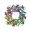
| Title | HCN2 CNBD in complex with inosine-3', 5'-cyclic monophosphate (cIMP) | ||||||
 Components Components | Potassium/sodium hyperpolarization-activated cyclic nucleotide-gated channel 2 | ||||||
 Keywords Keywords | TRANSPORT PROTEIN / protein-ligand complex / cycilc nucleotide binding domain / ion transport | ||||||
| Function / homology |  Function and homology information Function and homology informationHCN channels / cellular response to aldosterone / HCN channel complex / ammonium transmembrane transport / cellular response to cGMP / intracellularly cAMP-activated cation channel activity / sodium ion import across plasma membrane / voltage-gated sodium channel activity / regulation of membrane depolarization / potassium ion import across plasma membrane ...HCN channels / cellular response to aldosterone / HCN channel complex / ammonium transmembrane transport / cellular response to cGMP / intracellularly cAMP-activated cation channel activity / sodium ion import across plasma membrane / voltage-gated sodium channel activity / regulation of membrane depolarization / potassium ion import across plasma membrane / voltage-gated potassium channel activity / cAMP binding / somatodendritic compartment / potassium ion transmembrane transport / dendrite membrane / cellular response to cAMP / dendritic shaft / sodium ion transmembrane transport / PDZ domain binding / regulation of membrane potential / molecular adaptor activity / response to xenobiotic stimulus / axon / neuronal cell body / dendrite / protein-containing complex binding / membrane / identical protein binding / plasma membrane Similarity search - Function | ||||||
| Biological species |  | ||||||
| Method |  X-RAY DIFFRACTION / SYNCHROTRON / MOLECULAR REPLACEMENT / Resolution: 1.77 Å X-RAY DIFFRACTION / SYNCHROTRON / MOLECULAR REPLACEMENT / Resolution: 1.77 Å | ||||||
 Authors Authors | Ng, L.C.T. / Putrenko, I. / Baronas, V. / Van Petegem, F. / Accili, E.A. | ||||||
 Citation Citation | Journal: Structure / Year: 2016 Title: Cyclic Purine and Pyrimidine Nucleotides Bind to the HCN2 Ion Channel and Variably Promote C-Terminal Domain Interactions and Opening. Authors: Ng, L.C. / Putrenko, I. / Baronas, V. / Van Petegem, F. / Accili, E.A. | ||||||
| History |
|
- Structure visualization
| Structure viewer | Molecule: MolmilJmol/JSmol |
|---|
- Downloads & links
-Download
| PDBx/mmCIF format | 5khh.cif.gz | 61.2 KB | Display | PDBx/mmCIF format |
|---|---|---|---|---|
| PDB format | pdb5khh.ent.gz | 42.9 KB | Display | PDB format |
| PDBx/mmJSON format | 5khh.json.gz | Tree view | PDBx/mmJSON format | |
| Others |  Other downloads Other downloads |
-Validation report
| Arichive directory | https://data.pdbj.org/pub/pdb/validation_reports/kh/5khhftp://data.pdbj.org/pub/pdb/validation_reports/kh/5khh | HTTPS FTP |
|---|
-Related structure data
| Related structure data | 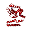 5khgC  5khiC 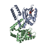 5khjC 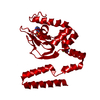 5khkC  1q5oS C: citing same article ( S: Starting model for refinement |
|---|---|
| Similar structure data |


-Links
 PDBj
PDBj


- Assembly
| Deposited unit | 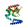
| ||||||||
|---|---|---|---|---|---|---|---|---|---|
| 1 | 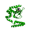
| ||||||||
| Unit cell |
|
-Components
| #1: Protein | Mass: 23883.330 Da / Num. of mol.: 1 / Fragment: UNP residues 443-643 Source method: isolated from a genetically manipulated source Source: (gene. exp.)  |
|---|---|
| #2: Chemical | ChemComp-6SW /   Mass: 330.191 Da / Num. of mol.: 1 / Source method: obtained synthetically / Formula: C10H11N4O7P Mass: 330.191 Da / Num. of mol.: 1 / Source method: obtained synthetically / Formula: C10H11N4O7P |
| #3: Water | ChemComp-HOH /  Mass: 18.015 Da / Num. of mol.: 165 / Source method: isolated from a natural source / Formula: H2O Mass: 18.015 Da / Num. of mol.: 165 / Source method: isolated from a natural source / Formula: H2O |
-Experimental details
-Experiment
| Experiment | Method: X-RAY DIFFRACTION / Number of used crystals: 1 |
|---|
- Sample preparation
| Crystal | Density Matthews: 2.91 Å3/Da / Density % sol: 57.72 % |
|---|---|
| Crystal grow | Temperature: 277 K / Method: vapor diffusion, hanging drop / pH: 5.5 Details: 200 mM NaCl, 0.1 mM sodium citrate pH 5.5, 14.5% PEG 400 |
-Data collection
| Diffraction | Mean temperature: 100 K | ||||||||||||||||||||||||||||||||||||||||||||||||||
|---|---|---|---|---|---|---|---|---|---|---|---|---|---|---|---|---|---|---|---|---|---|---|---|---|---|---|---|---|---|---|---|---|---|---|---|---|---|---|---|---|---|---|---|---|---|---|---|---|---|---|---|
| Diffraction source | Source: SYNCHROTRON / Site: APS  / Beamline: 23-ID-D / Wavelength: 0.97944 Å / Beamline: 23-ID-D / Wavelength: 0.97944 Å | ||||||||||||||||||||||||||||||||||||||||||||||||||
| Detector | Type: PSI PILATUS 6M / Detector: PIXEL / Date: Nov 30, 2012 / Details: 1000 um thick sensor | ||||||||||||||||||||||||||||||||||||||||||||||||||
| Radiation | Monochromator: Si(111) / Protocol: SINGLE WAVELENGTH / Monochromatic (M) / Laue (L): M / Scattering type: x-ray | ||||||||||||||||||||||||||||||||||||||||||||||||||
| Radiation wavelength | Wavelength: 0.97944 Å / Relative weight: 1 | ||||||||||||||||||||||||||||||||||||||||||||||||||
| Reflection | Resolution: 1.77→44.12 Å / Num. obs: 26044 / % possible obs: 97.5 % / Observed criterion σ(I): -3 / Redundancy: 9.83 % / Biso Wilson estimate: 42.756 Å2 / CC1/2: 0.998 / Rmerge(I) obs: 0.07 / Net I/σ(I): 19.06 | ||||||||||||||||||||||||||||||||||||||||||||||||||
| Reflection shell |
|
- Processing
| Software |
| ||||||||||||||||||||||||||||||||||||||||||||||||||||||||||||
|---|---|---|---|---|---|---|---|---|---|---|---|---|---|---|---|---|---|---|---|---|---|---|---|---|---|---|---|---|---|---|---|---|---|---|---|---|---|---|---|---|---|---|---|---|---|---|---|---|---|---|---|---|---|---|---|---|---|---|---|---|---|
| Refinement | Method to determine structure: MOLECULAR REPLACEMENT Starting model: 1Q5O Resolution: 1.77→44.12 Å / Cor.coef. Fo:Fc: 0.954 / Cor.coef. Fo:Fc free: 0.926 / SU B: 2.738 / SU ML: 0.086 / Cross valid method: THROUGHOUT / σ(F): 0 / ESU R: 0.127 / ESU R Free: 0.13 / Details: molecular replacement
| ||||||||||||||||||||||||||||||||||||||||||||||||||||||||||||
| Solvent computation | Ion probe radii: 0.8 Å / Shrinkage radii: 0.8 Å / VDW probe radii: 1.2 Å | ||||||||||||||||||||||||||||||||||||||||||||||||||||||||||||
| Displacement parameters | Biso max: 79.67 Å2 / Biso mean: 36.579 Å2 / Biso min: 21.85 Å2
| ||||||||||||||||||||||||||||||||||||||||||||||||||||||||||||
| Refinement step | Cycle: final / Resolution: 1.77→44.12 Å
| ||||||||||||||||||||||||||||||||||||||||||||||||||||||||||||
| Refine LS restraints |
| ||||||||||||||||||||||||||||||||||||||||||||||||||||||||||||
| LS refinement shell | Resolution: 1.771→1.817 Å / Total num. of bins used: 20
|
